Ultrasound imaging apparatus, image processing apparatus, image processing method, and computer program product

a technology of ultrasonic imaging and image processing, applied in ultrasonic/sonic/infrasonic diagnostics, instruments, tomography, etc., can solve problems such as difficulty in recognizing the direction in which the substance moves

Inactive Publication Date: 2010-09-02
TOSHIBA MEDICAL SYST CORP
View PDF7 Cites 28 Cited by
  • Summary
  • Abstract
  • Description
  • Claims
  • Application Information

AI Technical Summary

Problems solved by technology

However, when displaying an arbitrary cross section by color Doppler display by turning a three-dimensional image, the position of a probe is not recognized despite that the display is based on the position of the probe, it is difficult to recognize a direction in which the substance moves.

Method used


Image

  • Ultrasound imaging apparatus, image processing apparatus, image processing method, and computer program product
  • Ultrasound imaging apparatus, image processing apparatus, image processing method, and computer program product
  • Ultrasound imaging apparatus, image processing apparatus, image processing method, and computer program product

Examples


Embodiment Construction

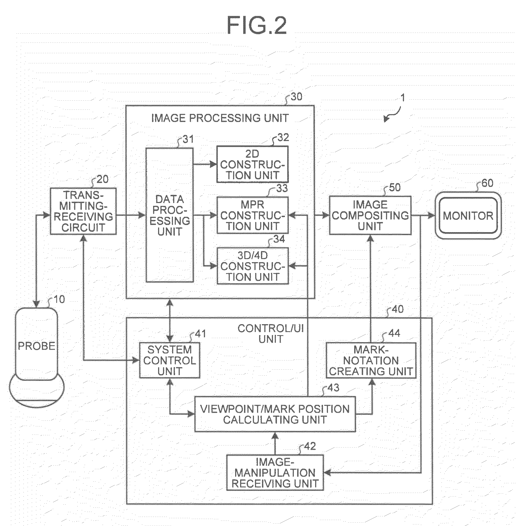
[0017]Exemplary embodiments of an ultrasound imaging apparatus, an image processing apparatus, an image processing method, and a computer program product according to the present invention will be explained below in detail with reference to the accompanying drawings.

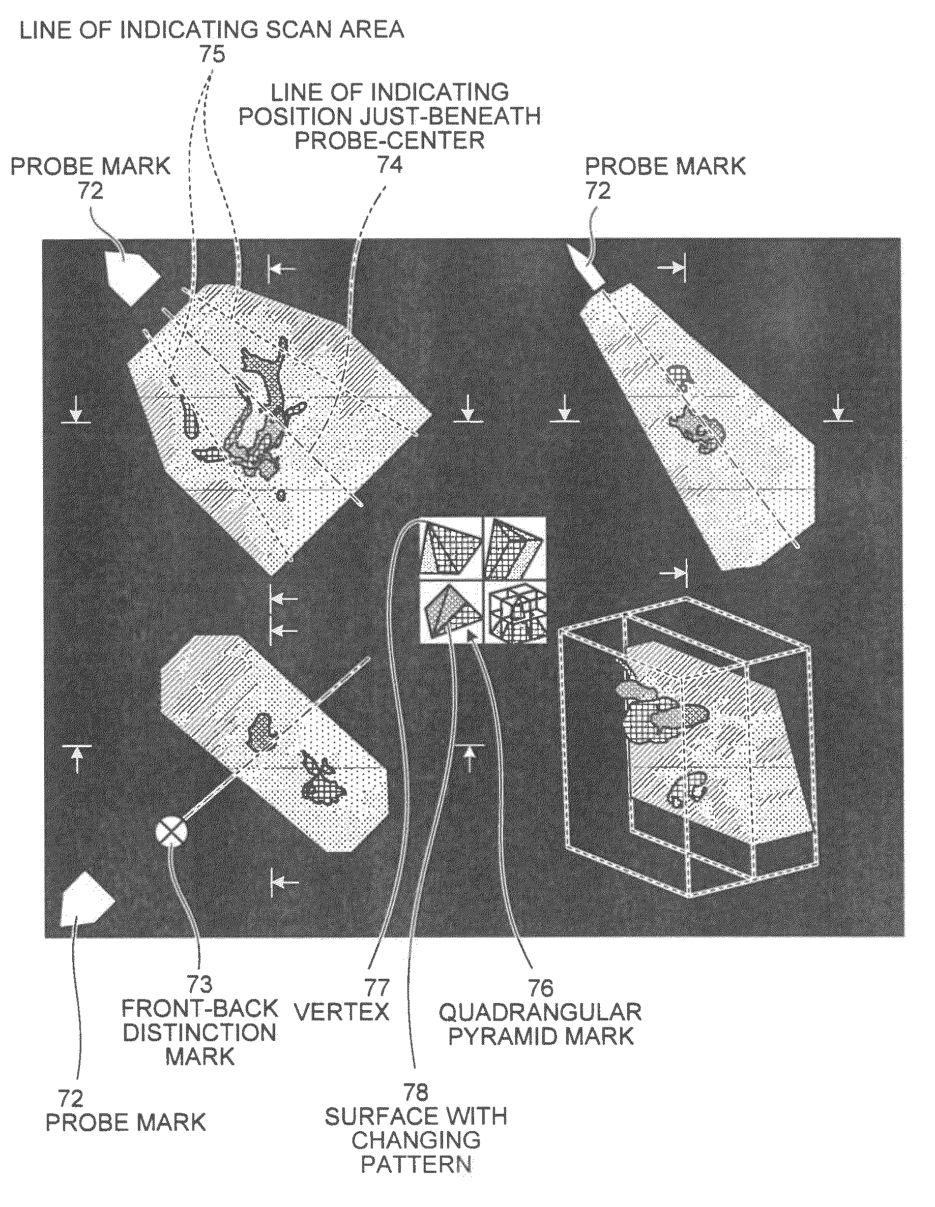
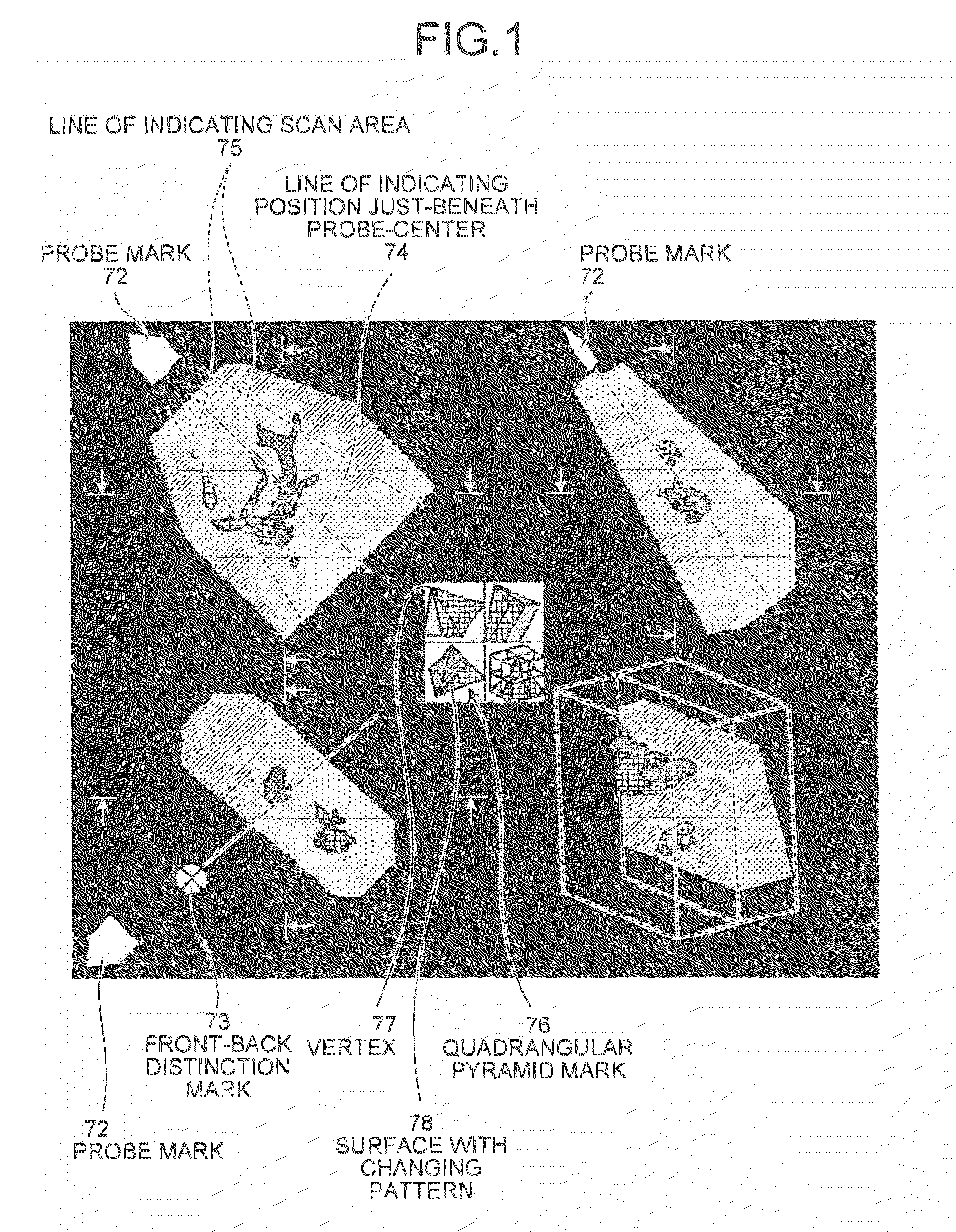
[0018]First of all, a Multi Planar Reconstruction (MPR) image and a three-dimensional image displayed by an ultrasound diagnosis apparatus according to an embodiment of the present invention are explained below. FIG. 1 is a schematic diagram that depicts MPR images and a three-dimensional image displayed by the ultrasound diagnosis apparatus according to the embodiment.

[0019]As shown in FIG. 1, the ultrasound diagnosis apparatus according to the embodiment displays a probe mark 72 that indicates a direction in which a probe is present on a scale of each color Doppler image displayed by MPR display. When the probe is positioned within a display area, the ultrasound diagnosis apparatus displays the probe mark 72 at the pos...


PUM


Abstract

An image-manipulation receiving unit receives an image manipulation by a user. A viewpoint/mark calculating unit calculates a viewpoint and a display position of a probe mark based on the image manipulation by the user. A mark-notation creating unit creates a probe mark, a front-back distinction mark, a line of indicating position just beneath probe center, a line of indicating scan area, and a quadrangular pyramid mark, as a mark. An image compositing unit then composites a color Doppler image with the marks, and displays them onto a monitor.

Description

CROSS-REFERENCE TO RELATED APPLICATIONS[0001]This application is based upon and claims the benefit of priority from the prior Japanese Patent Application No. 2009-047095, filed on Feb. 27, 2009; the entire contents of which are incorporated herein by reference.BACKGROUND OF THE INVENTION[0002]1. Field of the Invention[0003]The present invention relates to a technology for displaying an image taken by an ultrasound imaging apparatus, such as a color Doppler image.[0004]2. Description of the Related Art[0005]An ultrasound imaging apparatus is configured to display information about velocity, such as velocity in a blood vessel, in color as a color Doppler image (for example, see JP-A 2008-237759 (KOKAI)). Moreover, an ultrasound imaging apparatus is configured to display of a power component of a blood flow by using a three-dimensional image, and to display velocity information about an arbitrary cross section specified by a user by using a three-dimensional image as a color Doppler im...

Claims


Application Information

Patent Timeline
Patent Type & Authority Applications(United States)
IPC IPC(8): A61B8/14G06K9/00
CPCA61B8/06A61B8/13A61B8/42A61B8/463A61B8/483A61B8/488G06T2219/008G01S7/52074G01S7/52084G01S15/8979G01S15/8993G06T15/08G06T19/00G01S7/52073
Inventor HAMADA, KENJI
Owner TOSHIBA MEDICAL SYST CORP
Why Eureka
Try Eureka