Method and apparatus for enhanced in vivo MRI imaging

a mri imaging and in vivo technology, applied in the field of magnetic resonance imaging, can solve the problems of lack of specificity of ct in some situations, inability to blindly distinguish between inflammation and fibrosis, and inability to demonstrate the admixture of the two

Active Publication Date: 2010-09-09
BATTELLE MEMORIAL INST
View PDF4 Cites 6 Cited by
  • Summary
  • Abstract
  • Description
  • Claims
  • Application Information

AI Technical Summary

Benefits of technology

[0030]To assist one using the present invention in realizing the full benefits of the present invention, the invention preferably performs the additional step of highlighting the data points from the second set of measured data points that do not fall within the confidence region onto their location in an image of the second tissue. In this manner, the practitioner can readily identify the e

Problems solved by technology

The most routinely used modality for thoracic imaging is x-ray computed tomography (CT); however, CT lacks specificity in some situations.
However, results were generally bas

Method used


Image

  • Method and apparatus for enhanced in vivo MRI imaging
  • Method and apparatus for enhanced in vivo MRI imaging
  • Method and apparatus for enhanced in vivo MRI imaging

Examples


Embodiment Construction

[0041]For the purposes of promoting an understanding of the principles of the invention, reference will now be made to the embodiments illustrated in the drawings and specific language will be used to describe the same. It will nevertheless be understood that no limitations of the inventive scope is thereby intended, as the scope of this invention should be evaluated with reference to the claims appended hereto. Alterations and further modifications in the illustrated devices, and such further applications of the principles of the invention as illustrated herein are contemplated as would normally occur to one skilled in the art to which the invention relates.

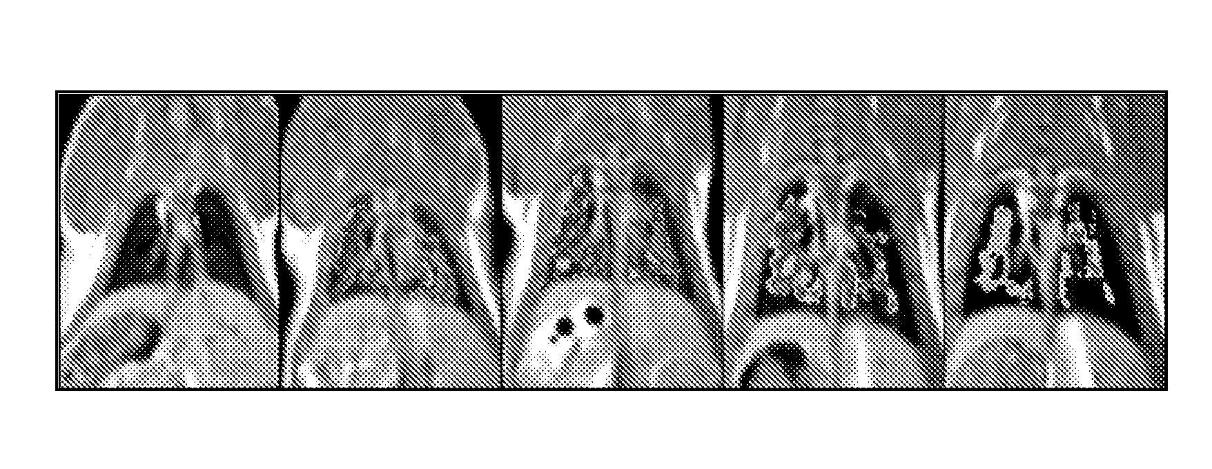
[0042]To demonstrate the present invention, a series of experiments were conducted to investigate the ability of the present invention to use proton (1H) magnetic resonance imaging (MRI) to distinguish between pulmonary inflammation and fibrosis. In these experiments, three groups of Sprague-Dawley rats (n=5) were instilled intr...


PUM


Abstract

A method and apparatus for detecting the presence of abnormal tissues of the present invention utilizing a magnetic resonance imaging system in communication with a computer. The apparatus is configured to define a confidence region using a probability based confidence interval calculation such as multivariate or bivariate analysis for at least two parameters in a normal tissue sample by making a magnetic resonance image, and then to evaluate a second sample by making a magnetic resonance image. Parameters detectable by magnetic resonance imaging 3 are selected from t1, t2, t2*, signal intensity, and diffusion.

Description

CROSS-REFERENCE TO RELATED APPLICATIONS[0001]This invention claims priority to Provisional Application Ser. No. 61 / 155,225 which was filed on Feb. 25, 2009.TECHNICAL FIELD[0002]This invention relates to magnetic resonance imaging (MRI). More specifically, this invention relates to improved techniques for distinguishing healthy tissues from diseased or injured tissues using MRI.BACKGROUND OF THE INVENTION[0003]For more than 20 years, researchers have been attempting to apply magnetic resonance imaging (MRI) to the characterization and diagnosis of pulmonary diseases. In part, this is motivated by the ongoing need for non-invasive methods to diagnose and stage interstitial diseases that challenge clinicians. Of particular interest in pulmonary MRI research is the potential to distinguish inflammation from fibrosis regionally and non-invasively. Although this is important for diagnosis and characterization of disease and disease activity, emphasis is often placed on assessing and diffe...

Claims


Application Information

Patent Timeline
IPC IPC(8): G01R33/44
CPCG01R33/50G01N24/08
Inventor JACOB, RICHARD E.AMIDAN, BRETTMINARD, KEVIN
Owner BATTELLE MEMORIAL INST
Why Eureka
Try Eureka