

Method for intrathecal administration of autologous stem cells in premature infants
a technology for autologous stem cells and premature infants, applied in the field of autologous stem cells for premature infants intrathecal administration, can solve the problems of lifelong seizures, interfere with normal neural pathways, and “healing” in a “non-functional” manner, and achieve the effects of minimizing the risk of autologous cells in this setting, minimizing the sequestration of pulmonary stem cells, and minimizing the potential value and limited toxicity
- Summary
- Abstract
- Description
- Claims
- Application Information
AI Technical Summary
Benefits of technology
Problems solved by technology
Method used
Image

Examples
Embodiment Construction
[0024]The present invention will be more fully understood and appreciated by reading the following Detailed Description in conjunction with the accompanying drawings, wherein like reference numerals refer to like components.
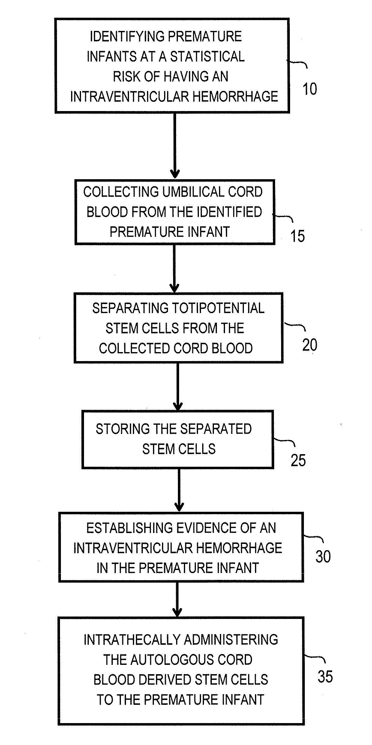
[0025]FIG. 3 is a flow chart illustrating steps involved in practicing a method for intrathecal administration of autologous cord blood derived stem cells in premature infants sustaining intraventricular hemorrhages, according to an embodiment of the present invention. The method, or therapeutic protocol, can include one or more of the following steps: identifying premature infants at a statistical risk of having an intraventricular hemorrhage 10, collecting umbilical cord blood from the identified premature infant 15, separating totipotential stem cells (having the ability to proliferate and differentiate as neural stem cells) from the collected cord blood 20, storing the separated stem cells 25, establishing evidence of an intraventricular hemorrhage (preferabl...
PUM
 Abstract
Description
Claims
Application Information