Method and system for blood vessel segmentation and classification

a blood vessel and segmentation technology, applied in the field of medical imaging segmentation, can solve the problems of easy error and tedious medical image inspection

Active Publication Date: 2010-11-25
PHILIPS MEDICAL SYST TECH
View PDF11 Cites 101 Cited by
  • Summary
  • Abstract
  • Description
  • Claims
  • Application Information

AI Technical Summary

Benefits of technology

[0065]For example, hardware for performing selected tasks according to embodiments of the invention could be implemented as a chip or a circuit. As software, selected tasks according to embodiments of the invention could be implemented as a plurality of software instructions being executed by a computer using any suitable operating system. In an exemplary embodiment of the invention, one or more tasks according to exemplary embodiments of method and/or system as described he...

Problems solved by technology

Thus, inspection of medical ima...

Method used


Image

  • Method and system for blood vessel segmentation and classification
  • Method and system for blood vessel segmentation and classification
  • Method and system for blood vessel segmentation and classification

Examples


Embodiment Construction

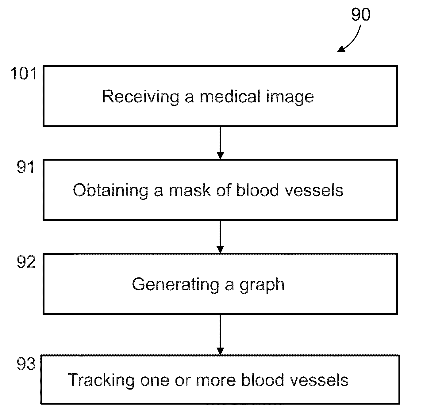
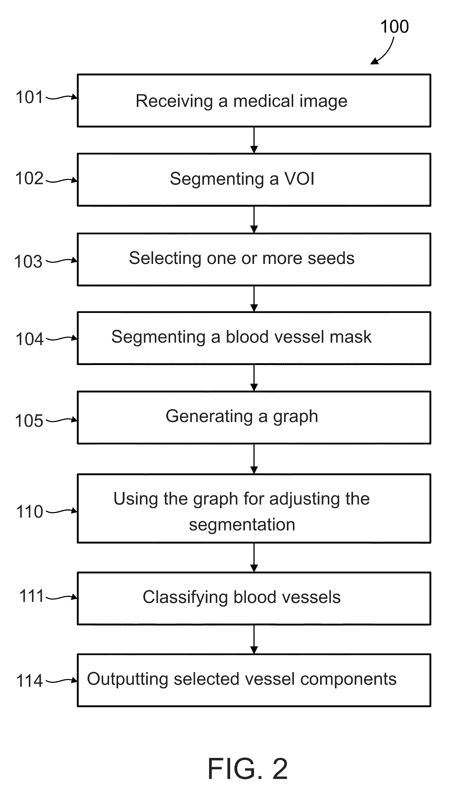
[0079]The present invention, in some embodiments thereof, relates to medical imaging segmentation and, more particularly, but not exclusively, to segmentation of blood vessels and networks of blood vessels in medical imaging applications.

[0080]According to some embodiments of the present invention there is provided a method for analyzing the structure of networks of blood vessels, such as the coronary arteries, the hepatic artery tree, hepatic vein tree, portal vein, and / or portal vein branches, in a medical image, such as a CT-angiography study, for example a CT study of the heart during a selected phase representing a predetermined period of a beat of the heart. The method is based on analyzing the topology of the network in order to find one or more target blood vessels in a selected network of blood vessels using a graph that maps a plurality of paths in the network. The graph, which may have both directed and undirected edges to represent knowledge about the direction of flow i...


PUM


Abstract

A method of analyzing structure of a network of vessels in a medical image, comprising:
    • receiving the medical image depicting the network of vessels;
    • obtaining a mask of the network of vessels in the image; and
    • generating a non-forest graph mapping a plurality of paths of vessels in the network to directed paths in the graph, with each edge in the graph either directed to indicate a known direction of flow in the corresponding vessel, or undirected to indicate a lack of knowledge of direction of flow in the corresponding vessel, and with all directed edges in a path directed in a same direction as the path.

Description

RELATED APPLICATIONS[0001]This application claims benefit under 35 USC 119(e) of U.S. Provisional Patent Application No. 61 / 179,386 filed on May 19, 2009. The contents of that application are incorporated by reference as if fully set forth herein.FIELD AND BACKGROUND OF THE INVENTION[0002]The present invention, in some embodiments thereof, relates to medical imaging segmentation and, more particularly, but not exclusively, to segmentation of blood vessels and blood vessel networks in medical imaging applications.[0003]Medical images are images of a human subject that are analyzed for the purposes of biological and medical research, diagnosing and treating disease, injury and birth defects. Commonly, medical images involve modalities that are designed for capturing data for imaging internal organs and tissues in a non-invasive manner. Examples of such modalities include computed tomography (CT), magnetic resonance imaging (MRI), positron emission tomography (PET), ultrasound, fluoros...

Claims


Application Information

Patent Timeline
IPC IPC(8): G06K9/00
CPCG06K2009/00932G06T7/0081G06T2207/30101G06T2207/10072G06T7/0093G06T7/11G06T7/162G06V40/14
Inventor OSTROVSKY-BERMAN, YARONPORAT, HADARGAZIT, TIFERET AHAVAHDROR, ORANIT
Owner PHILIPS MEDICAL SYST TECH
Why Eureka
Try Eureka