Method and Apparatus for Determining Angulation of C-Arm Image Acquisition System for Aortic Valve Implantation

a technology of aortic valve and image acquisition system, which is applied in the field of medical imaging of the heart, can solve the problems of large amount of contrast agent and time-consuming, and achieve the effect of limiting the exposure of patients to contrast agen

Inactive Publication Date: 2011-03-03
SIEMENS AG
View PDF9 Cites 28 Cited by
  • Summary
  • Abstract
  • Description
  • Claims
  • Application Information

AI Technical Summary

Problems solved by technology

Otherwise, the physician may position the valve prosthesis such that the valve prosthesis accidently closes the coronary arteries.
Accordingly, a challenge in conventional valve implantation is for the physician to find a good perpendicular view for the C-arm X-ray system.
Accordingly, this selection process typically requires a large amount of contrast agent and is time consuming.

Method used


Image

  • Method and Apparatus for Determining Angulation of C-Arm Image Acquisition System for Aortic Valve Implantation
  • Method and Apparatus for Determining Angulation of C-Arm Image Acquisition System for Aortic Valve Implantation
  • Method and Apparatus for Determining Angulation of C-Arm Image Acquisition System for Aortic Valve Implantation

Examples


Embodiment Construction

[0015]The present invention is directed to a method and system for determining an optimal angulation of a C-arm image acquisition system for aortic valve implantation using 3D medical images, such as DynaCT images, cardiac CT images, and cardiac MR images. Embodiments of the present invention are described herein to give a visual understanding of the method for determining an optimal angulation. A digital image is often composed of digital representations of one or more objects (or shapes). The digital representation of an object is often described herein in terms of identifying and manipulating the objects. Such manipulations are virtual manipulations accomplished in the memory or other circuitry / hardware of a computer system. Accordingly, it is to be understood that embodiments of the present invention may be performed within a computer system using data stored within the computer system.

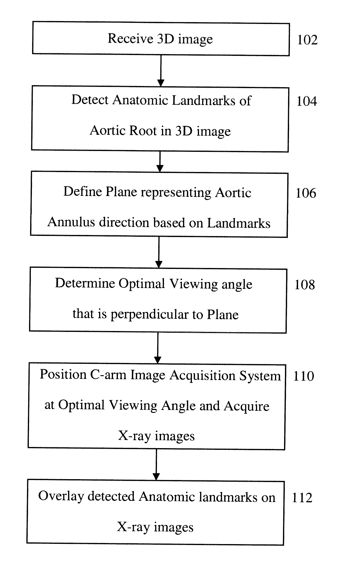
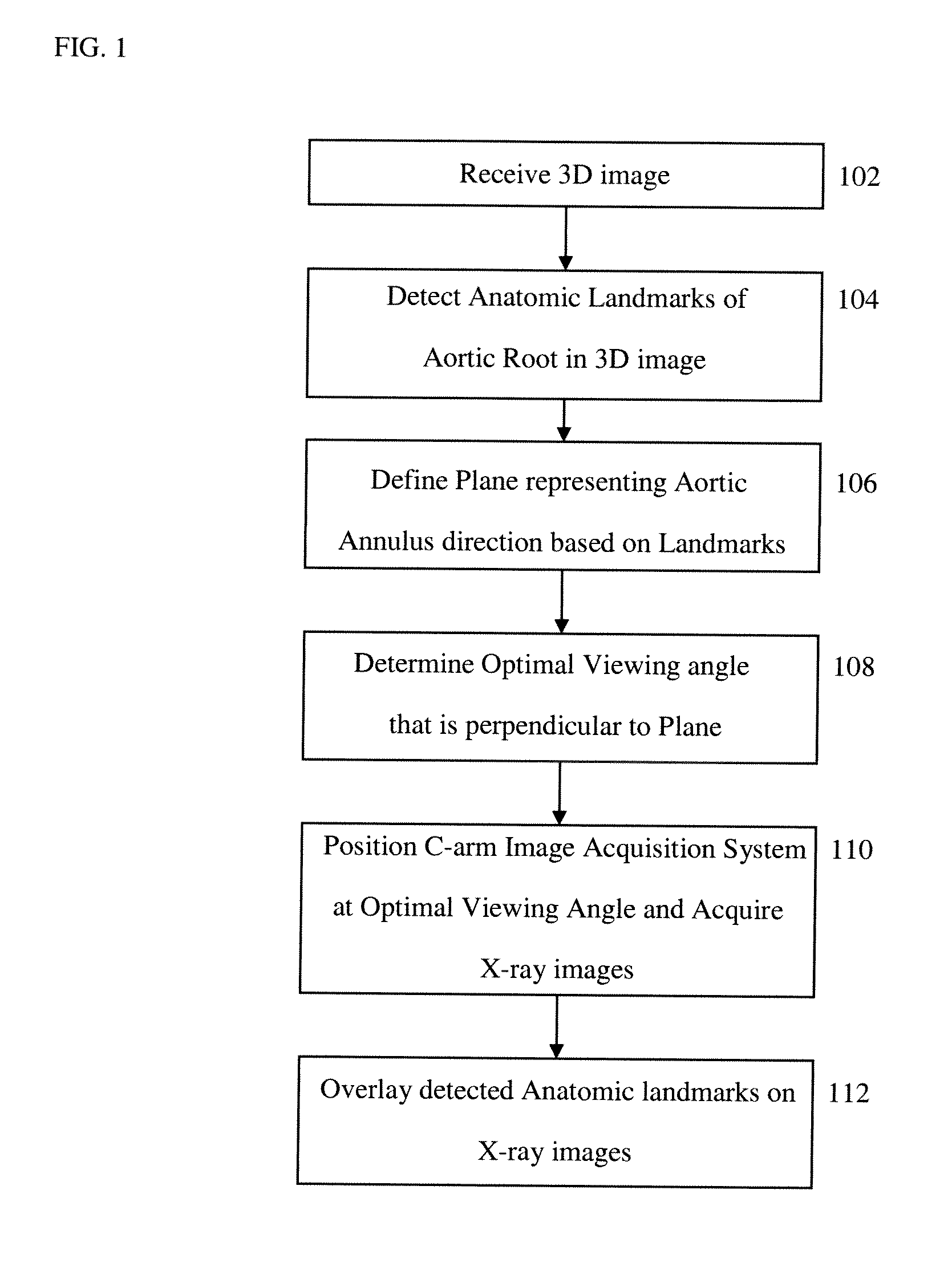
[0016]FIG. 1 illustrates a method for determining an optimal angulation of a C-arm image acqui...


PUM


Abstract

A method and system for determining an angulation of a C-arm image acquisition system for aortic valve implantation is disclosed. One or more landmarks of the aortic root is detected in a 3D image. A plane representing an aortic annulus direction is defined in the 3D image based on the detected anatomic landmarks. A viewing angle is determined that is perpendicular to the defined plane.

Description

[0001]This application claims the benefit of U.S. Provisional Application No. 61 / 237,733, filed Aug. 28, 2009, the disclosure of which is herein incorporated by reference.BACKGROUND OF THE INVENTION[0002]The present invention relates to medical imaging of the heart, and more particularly, to using medical images of the heart to determine an angulation of a C-arm image acquisition system for aortic valve implantation.[0003]Aortic valve disease is the most common valvular disease in developed countries, and has the second highest incidence among congenital valvular defects. Implantation of an artificial valve (i.e., valve prosthesis) is often necessary to replace a damaged natural valve. In minimally invasive valve implantations, a valve prosthesis is inserted via a catheter and X-ray imaging is used to support a physician in positioning and deployment of the valve prosthesis. In particular, during a valve implantation surgery, 2D fluoroscopic images (X-ray images) are often captured ...

Claims


Application Information

Patent Timeline
Patent Type & Authority Applications(United States)
IPC IPC(8): G06K9/00
CPCG06T7/0042G06T2207/30101G06T2207/30052G06T2207/10116G06T7/73
Inventor LIAO, RUIZHENG, YEFENGJOHN, MATTHIASNOETTLING, ALOISBOESE, JAN
Owner SIEMENS AG
Why Eureka
Try Eureka