Use of Epineural Sheath Grafts for Neural Regeneration and Protection

a technology of epineural sheath and grafts, which is applied in the field of neural regeneration and protection, can solve the problems of significant gaps, optimal functional outcome, and major challenges in the surgical management of nerve defects, and achieve the effect of minimising inflammatory reactions

Active Publication Date: 2011-04-14
THE CLEVELAND CLINIC FOUND
View PDF24 Cites 29 Cited by
  • Summary
  • Abstract
  • Description
  • Claims
  • Application Information

AI Technical Summary

Benefits of technology

[0005]Described herein is conduit material that causes minimal inflammatory reaction, and can serve as a structural guide for regenerating, or as a shield for protecting, nerve tissue (e.g., axons). Thus, the invention is directed to methods of treating an injury to a (one or more) nerve or protecting a nerve in an individual in need thereof. The methods employ all or a portion of an isolated, naturally occurring epineural sheath, and can be used, for example, to regenerate nerve tissue in an individual in need thereof.

Problems solved by technology

Severe nerve injuries may result in significant gaps and surgical management of nerve defects is still a major challenge.
Currently, the microsurgical techniques used for the repair of peripheral nerve defects do not always result in optimal functional outcome.
There is also a limited source of donor nerves available that makes reconstruction of large nerve defects difficult.
Nerve allografts have been studied experimentally and used clinically, however the major disadvantage is the need for immunosuppression to prevent rejection (Mackinnon, S E., Plast. Reconstr. Surg. 107:1419-1429 (2001); Fox, I K, Muscle Nerve, 31:59-69 (2005)).
Therefore nerve allografts are used only in selected cases and are not the primary choice of peripheral nerve repair in clinical practice.
These conduits are used as tubulized chambers and present with different advantages, but also disadvantages such as inflammation, foreign body reaction, compression, and toxicity of degradation products.

Method used


Image

  • Use of Epineural Sheath Grafts for Neural Regeneration and Protection
  • Use of Epineural Sheath Grafts for Neural Regeneration and Protection
  • Use of Epineural Sheath Grafts for Neural Regeneration and Protection

Examples


example 1

Repair of Peripheral Nerve Defects with Epineural Sheath Graft

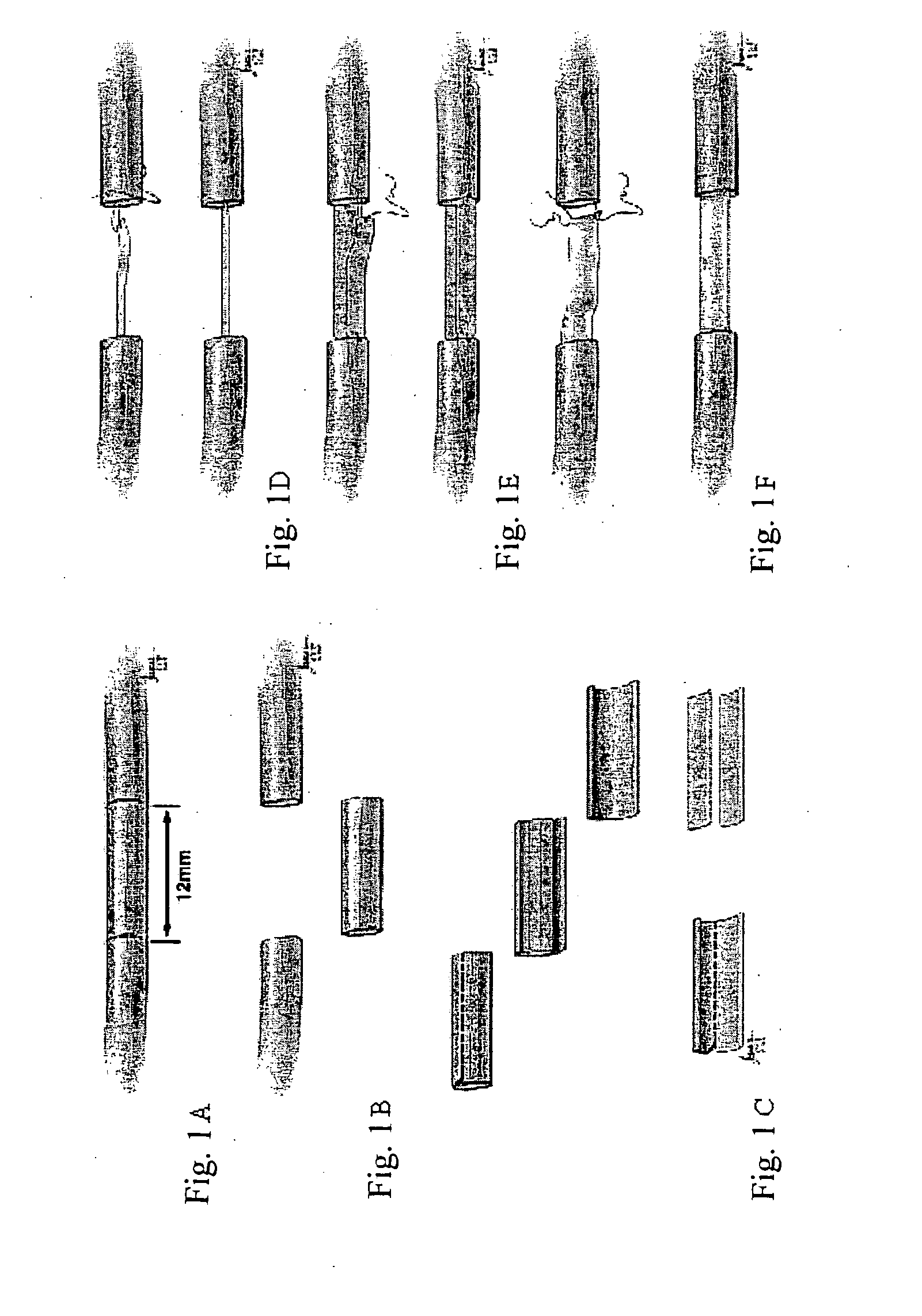
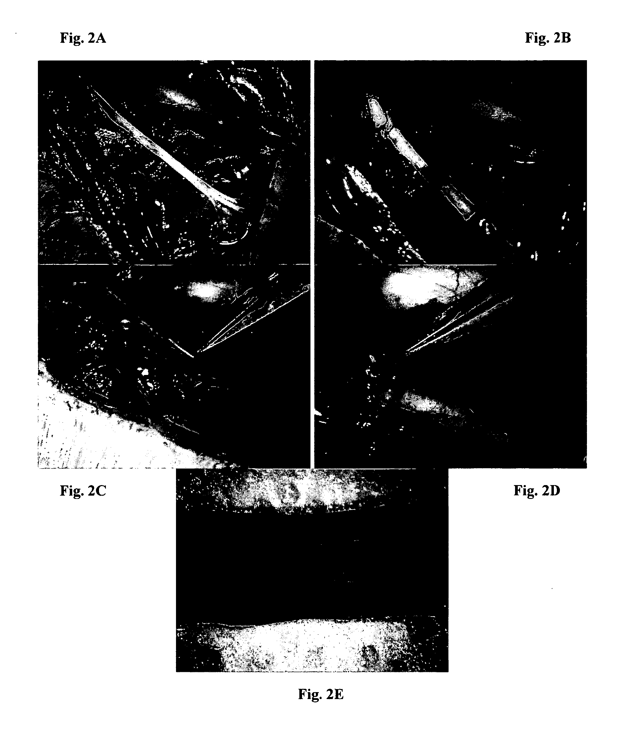
[0087]In this study, the potential of using detubulized flat epineural sheath strip for bridging nerve gaps as an alternative to nerve autografting technique was investigated. Nerve gaps were created by removing a 1.2 cm segment of the right sciatic nerves. The epineurium of the removed segment was incised longitudinally and by removing the fascicules, a flat rectangular shaped epineural sheath was obtained. The groups (6 rats of each) repaired with one strip, two-strip and full epineural sheath grafts were compared with the animals repaired with autografts and the untreated groups at 12th week. Toe-spread was better in rats repaired with full sheath grafts and conventional nerve grafts compared to single strip graft at 12th week. Somatosensory evoked potential evaluation demonstrated no significant difference in latencies between conventional and epineural sheath groups. Histomorphometric results of the sections from the...

example 2

Enhancement of Neural Regeneration of Peripheral Nerve Defects by Epineural Tube Graft Enriched with Donor Derived Bone Marrow Stromal Cells

[0113]Nerve grafting has been the most widely accepted surgical intervention for the repair of peripheral nerve defects. Traditionally however, such interventions have been associated with varying degrees of donor site morbidity. In the search for alternative methods of peripheral nerve repair, the use of biological and artificial materials for the creation of nerve conduits has been investigated. This study described herein was performed to assess the effects of bone marrow stromal cells (BMSC) in nerve gaps repaired with isogenic, isolated epineural tubes filled with isogenic and allogenic BMSCs.

Methods

[0114]A total of 54 isolated epineural tubes were harvested. Access to the peripheral nerve e.g. sciatic nerve was made by skin incision and subcutaneous tissue dissection down to the anatomical location of the nerve. At this level the sciatic n...

example 3

Immunostaining of the Epineural Tube

Methods:

[0121]The sciatic nerves were harvested from naïve Lewis rats (Lew RT11). The fascicles were removed using fascicle-stripper device. After fascicle removal empty epineural tube was created. This tube was next evaluated for expression of nerve factors involved in nerve growth and regeneration and was evaluated by monoclonal antibodies and immunofluorescence technique.

[0122]Next the cross-sections and longitudinal sections were prepared from whole tubes as well as from tubes split opened and let flat for the testing.

[0123]These freshly dissected nerve tubes from naïve Lewis rat were snap-frozen in liquid nitrogen. Prior to immunostaining tissue slides were cut for 5 μm slides and fixed for 10 min. in acetone. Next, sections were rinsed in TBS (Dako) buffer and incubated with mouse anti-rat NGF (H-20), GFAP (2E1), VEGF (C1) (Santa Cruz Biotechnology, Inc), S100 (clone 4C4.9) (LabVision), MHC class II (clone OX-17) and Laminin B (clone D18-2.2...


PUM


Abstract

Described herein is conduit material that causes minimal inflammatory reaction, and serves as a structural guide for regenerating nerve tissue (e.g., axons). Thus, the invention is directed to methods of treating a nerve injury in an individual in need thereof. The methods employ an isolated, naturally occurring epineural sheath, and can be used, for example, to regenerate nerve tissue in an individual in need thereof. Also provided herein is a device for harvesting an epineural sheath.

Description

RELATED APPLICATION(S)[0001]This application claims the benefit of U.S. Provisional Application No. 61 / 123,026, filed on Apr. 4, 2008. The entire teachings of the above application(s) are incorporated herein by reference.BACKGROUND OF THE INVENTION[0002]Severe nerve injuries may result in significant gaps and surgical management of nerve defects is still a major challenge. When direct repair of the injured nerve with epineural sutures is impossible, the defect between the nerve stumps has to be bridged by a conduit of some kind, which will facilitate axonal regeneration towards the distal nerve stump. Currently, the microsurgical techniques used for the repair of peripheral nerve defects do not always result in optimal functional outcome. The most reliable and widely used method is bridging the defect by an autograft, which is considered the gold standard for nerve gap repair (Watchmaker, GP et al., Clin. Plast. Surg, 24:63-73 (1997); Siemionow, M., et al., Ann. Plast. Surg., 52:72-...

Claims


Application Information

Patent Timeline
Patent Type & Authority Applications(United States)
IPC IPC(8): A61F2/02A61B17/32
CPCA61B17/1128A61B17/32002A61B2017/320064A61B2017/00969A61B2017/320044A61B2017/00349
Inventor SIEMIONOW, MARIASIEMIONOW, KRZYSZTOF
Owner THE CLEVELAND CLINIC FOUND
Why Eureka
Try Eureka