Systems and methods for measurement of objects of interest in medical images

a technology of objects and images, applied in image analysis, image enhancement, instruments, etc., can solve the problems of increasing the work load of the radiologist, slowing down the workflow, and consuming tim

Active Publication Date: 2011-05-26
FUJIFILM CORP
View PDF10 Cites 71 Cited by
  • Summary
  • Abstract
  • Description
  • Claims
  • Application Information

AI Technical Summary

Benefits of technology

[0010]Various embodiments of the invention relate to systems and methods for the measurement and display of attributes of an object of interest in a medical image. An object of interest is identified, such as a lesion or cluster of lesions in a breast area on a mammography image. At least one attribute of the lesion is then automatically measured, such as the area of the lesion, the width and height of a cluster of lesions, the number of lesions in a cluster, or the distance from one or more lesions to an anatomical feature such as the nipple, skin line or chest wall. The measurements are then displayed to a user, for example by mapping the measurements onto the mammography image. Additionally, anatomical zones, such as standard, quadrant and clock zones of the breast area may be determined and mapped onto the mammography image to display the location of the lesion as it corresponds to the zones. The measurements and the anatomical zone information can also be recorded and put into document files, such as a word processing template for a mammography reading report, which can be printed out later for viewing.

Problems solved by technology

However, these attributes are measured manually by the radiologist, which slows down the workflow and increases the work load of the radiologist.
The process is time consuming.

Method used


Image

  • Systems and methods for measurement of objects of interest in medical images
  • Systems and methods for measurement of objects of interest in medical images
  • Systems and methods for measurement of objects of interest in medical images

Examples


Embodiment Construction

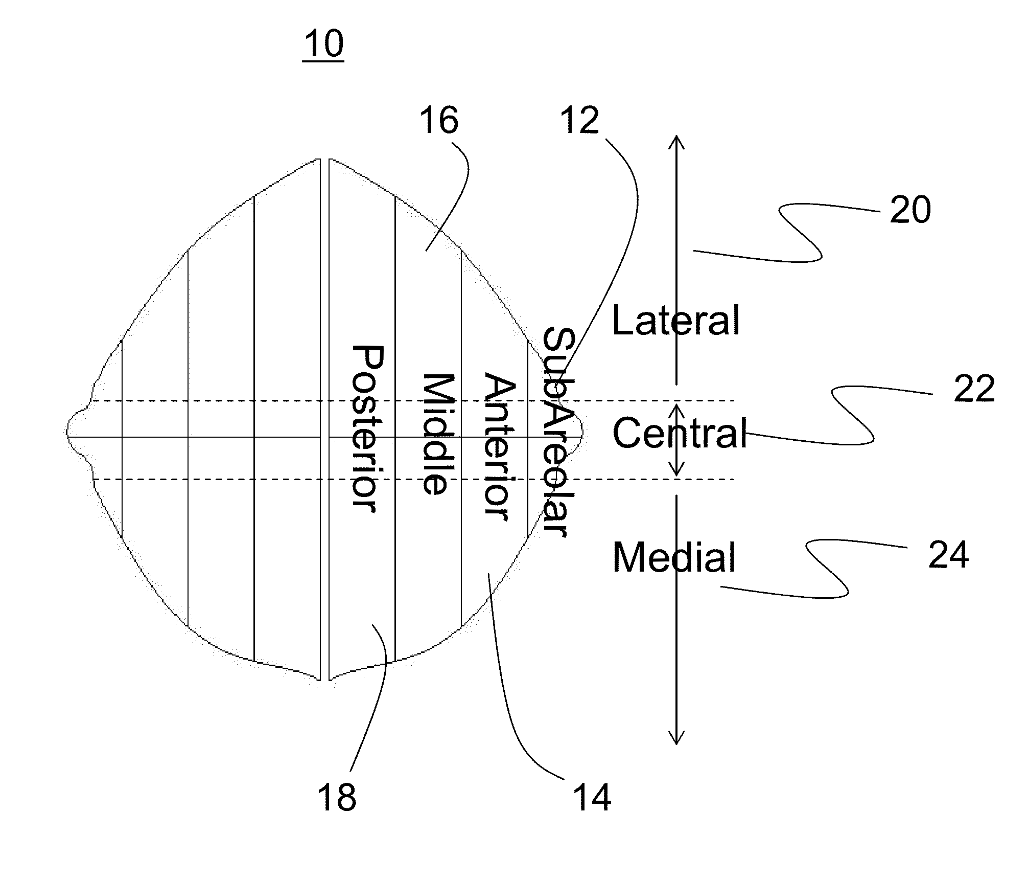
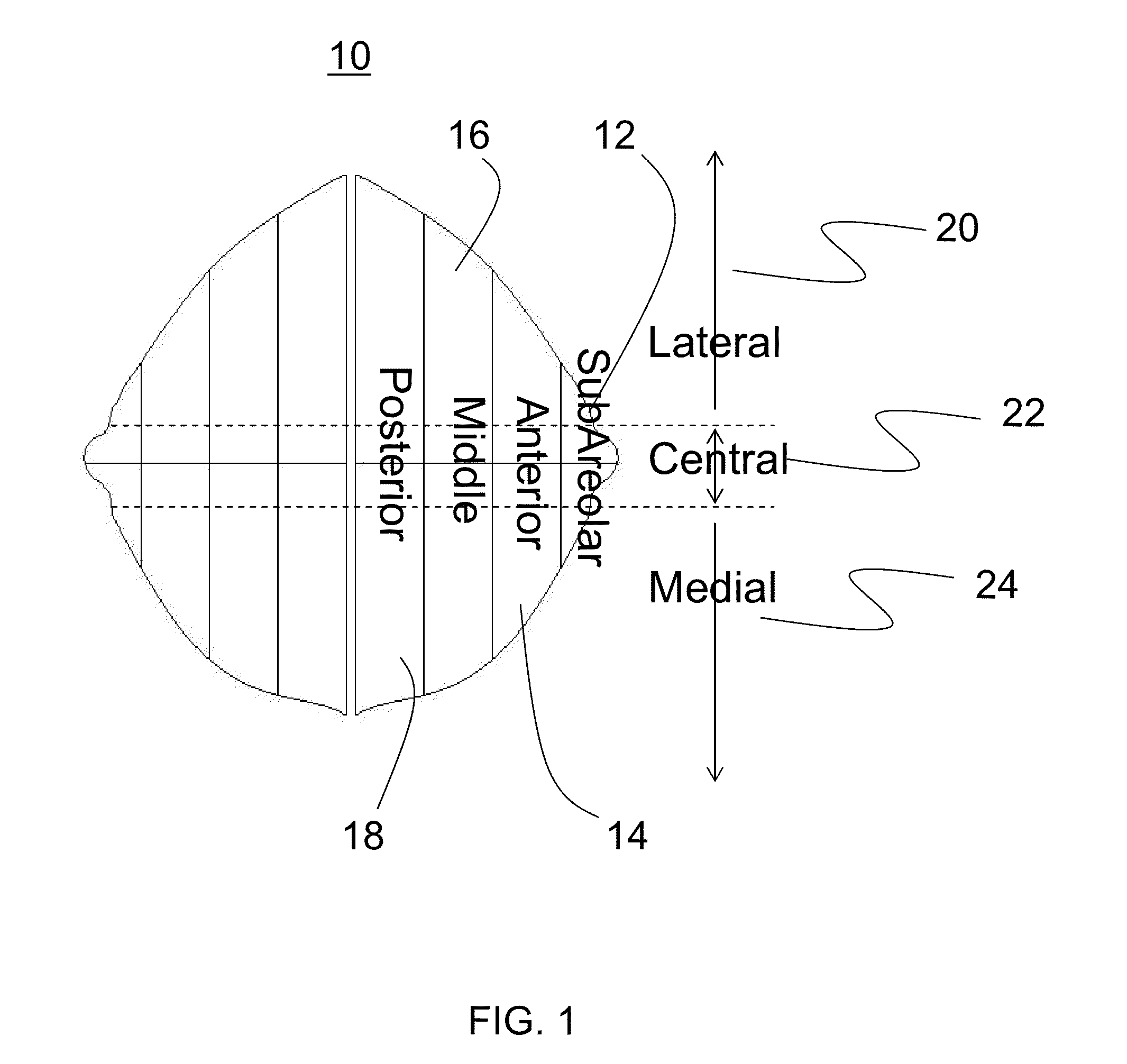
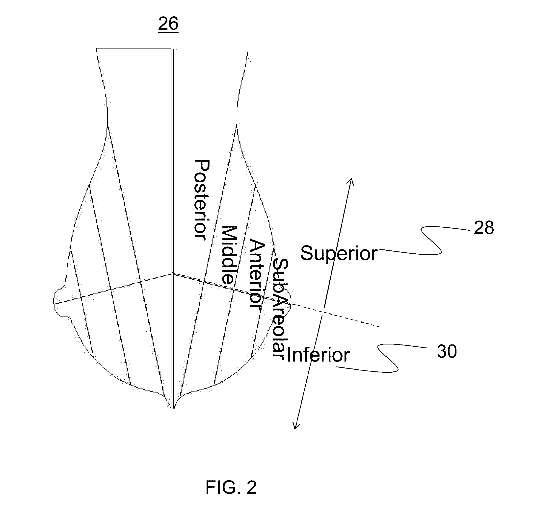
[0060]In the following detailed description, reference will be made to the accompanying drawing(s), in which identical functional elements are designated with like numerals. The aforementioned accompanying drawings show by way of illustration and not by way of limitation, specific embodiments and implementations consistent with principles of the present invention. These implementations are described in sufficient detail to enable those skilled in the art to practice the invention and it is to be understood that other implementations may be utilized and that structural changes and / or substitutions of various elements may be made without departing from the scope and spirit of present invention. The following detailed description is, therefore, not to be construed in a limited sense. Additionally, the various embodiments of the invention as described may be implemented in the form of software running on a general purpose computer, in the form of a specialized hardware, or combination o...


PUM


Abstract

A system and method are provided for the measurement and display of attributes of an object of interest in a medical image. An object of interest is identified, such as a lesion or cluster of lesions in a breast area on a mammography image. At least one attribute of the lesion is then automatically measured, such as the area of the lesion, the width and height of a cluster of lesions, the number of lesions in a cluster, or the distance from one or more lesions to an anatomical feature such as the nipple, skin line or chest wall. The measurements are then displayed to a user, for example by displaying the measurements on the mammography image. Additionally, anatomical zones, such as standard, quadrant and clock zones of the breast area may be determined and displayed on the mammography image or diagram to display the location of the lesion as it corresponds to the zones.

Description

BACKGROUND[0001]1. Field[0002]The systems and methods described below relate to the measurement and display of objects of interest in a medical image, and more specifically to automatic measurement and display of attributes of lesions in mammography images.[0003]2. Background[0004]Medical imaging is the field of creating images of the human body for medical purposes, such as diagnosing or examining disease or other physiological anomalies. Numerous types of image modalities produce medical images, such as magnetic resonance imaging (MRI), radiography (x-rays), computed tomography (CT), ultrasound (US) and others. In medical imaging, an object of interest is usually selected pertaining to an area of the human body, such as the head, heart or chest.[0005]One type of medical imaging is mammography, which is the examination of a medical image of the human breast. Mammography is used to detect breast cancer by examining the breast tissue for abnormalities such as lesions that represent m...

Claims


Application Information

Patent Timeline
Patent Type & Authority Applications(United States)
IPC IPC(8): G06K9/00G09G5/00
CPCG06T7/602G06T2207/30068G06T2207/10116G06T7/62
Inventor NIE, YAOSHI, CHAOMAJDI-NASAB, NARIMANHASEGAWA, AKIRA
Owner FUJIFILM CORP
Why Eureka
Try Eureka