Imaging System for Following a Surgical Tool in an Operation Field

a surgical tool and imaging system technology, applied in the field of medical imaging, can solve the problems of cumbersome implementation of existing solutions, time-consuming, and insufficient reliability of images using pre-operative data

Active Publication Date: 2011-06-02
UNIV GRENOBLE ALPES +1
View PDF20 Cites 19 Cited by
  • Summary
  • Abstract
  • Description
  • Claims
  • Application Information

AI Technical Summary

Problems solved by technology

However, existing solutions are relatively cumbersome to implement.
In addition, the images using pre-operative data are not fully reliable since there morphological changes may occur between the acquisition of this data and surgery (whether changes with respect to the inter-positioning of the organs and even changes in shape).
However, said solution is time-consuming since processing of the data is complex which means that viewing of the enhanced images cannot be made in real time.
However, said solution cannot be implemented under any surgical conditions since it must be possible to visualize both imaging devices.
In addition, said system is relatively voluminous which is a major drawback regarding required operating room space.

Method used


Image

  • Imaging System for Following a Surgical Tool in an Operation Field
  • Imaging System for Following a Surgical Tool in an Operation Field
  • Imaging System for Following a Surgical Tool in an Operation Field

Examples


Embodiment Construction

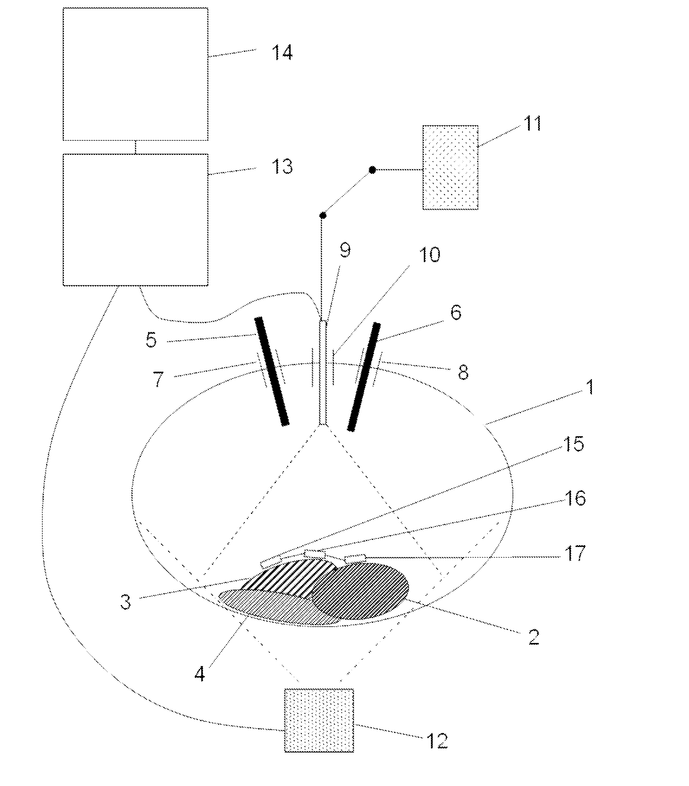
[0043]FIG. 1 schematically illustrates the imaging system for the monitoring of surgical instruments in an operative site inside a volume 1 of the body of a patient under endoscopic surgery. This volume 1, or volumetric region, forms a cavity 1, this cavity being natural or artificial created in this case by injection of air into the volume.

[0044]As schematically depicted in FIG. 1, one or more incisions are made in the cavity 1, these incisions being used to insert surgical instruments (5, 6). These incisions are made using trocars (7, 8) which are inserted through the wall forming the cavity 1 and which are used as portals for the instruments to be inserted inside the cavity 1.

[0045]During endoscopic surgery, it is essential for the surgeon to have a proper view of the instruments (5, 6) in relation to the organs (2, 3, 4) present in the operative site.

[0046]For this purpose, the imaging system comprises firstly conventional photosensitive imaging means inserted in the cavity 1 vi...


PUM


Abstract

The invention concerns an imaging system to monitor at least one surgical instrument in an operative site inside a volume of the body of an animal, comprising:
    • at least one endoscopic camera to obtain endoscopic data on the operative site,
    • at least one ultrasound imaging device to obtain ultrasound data on the operative site,
    • a processing device to process the endoscopic and ultrasound data,
characterized in that it further comprises at least three markers intended to be positioned in the operative site, said markers being mobile relative to the instrument, each marker being adapted to be detected both by the endoscopic camera and by the ultrasound imaging device, so as to permit cross-mapping of the endoscopic and ultrasound data by the processing means.

Description

FIELD OF THE INVENTION[0001]The present invention relates to the field of medical imaging, more particularly to the monitoring of a surgical instrument in an operative site.STATE OF THE ART[0002]Endoscopic surgery requires a high-performance imaging system for visualization of the operative site, to assist the surgeon during surgery.[0003]A certain number of solutions have been developed to improve the viewing offered by the image of a conventional endoscopic camera.[0004]Some systems use images of the operative site acquired before surgery by any system of CT-scan, magnetic resonance imaging, or stereoscopic camera type. Pre-operative images can effectively be used to segment the 3D volume acquired before surgery to isolate the organs of interest, and to relate them with the per-operative imaging system generally consisting of an endoscopic camera. The merging of the 3D images acquired before surgery with the endoscopic images acquired during surgery provide the surgeon with improv...

Claims


Application Information

Patent Timeline
Patent Type & Authority Applications(United States)
IPC IPC(8): A61B8/14A61B1/00
CPCA61B19/52A61B2019/5231A61B2019/5278A61B2019/5289A61B2019/5495A61B2019/5425A61B2019/5429A61B2019/5437A61B2019/5445A61B2019/5408A61B90/36A61B2090/364A61B2090/373A61B2090/3782A61B2090/3908A61B2090/3925A61B2090/3929A61B2090/3937A61B2090/3945A61B2090/3995
Inventor CINQUIN, PHILIPPEMOZER, PIERREVOROS, SANDRINELONG, JEAN-ALEXANDREDUCHATEAU, JOSSELINMOREAU GAUDRY, ALEXANDREVIDAL, CLEMENTHENRI, PATRICK
Owner UNIV GRENOBLE ALPES
Why Eureka
Try Eureka