Method and system for lesion segmentation

Inactive Publication Date: 2011-06-30
IBBT +3
View PDF8 Cites 44 Cited by
  • Summary
  • Abstract
  • Description
  • Claims
  • Application Information

AI Technical Summary

Problems solved by technology

Needless to say, this is a time-consuming task, requiring strict attentiveness to avoid medical errors.
The situation becomes even worse when several longitudinal studies have to be assessed for follow-up after chemotherapy or radiotherapy.
Such problems occur in studies involving abdominal organs (e.g., liver, kidney, pancreas, spleen, .
However, this approach of first segmenting each phase and then reconciling the segmentations in a registration suffers fr

Method used


Image

  • Method and system for lesion segmentation
  • Method and system for lesion segmentation
  • Method and system for lesion segmentation

Examples


example

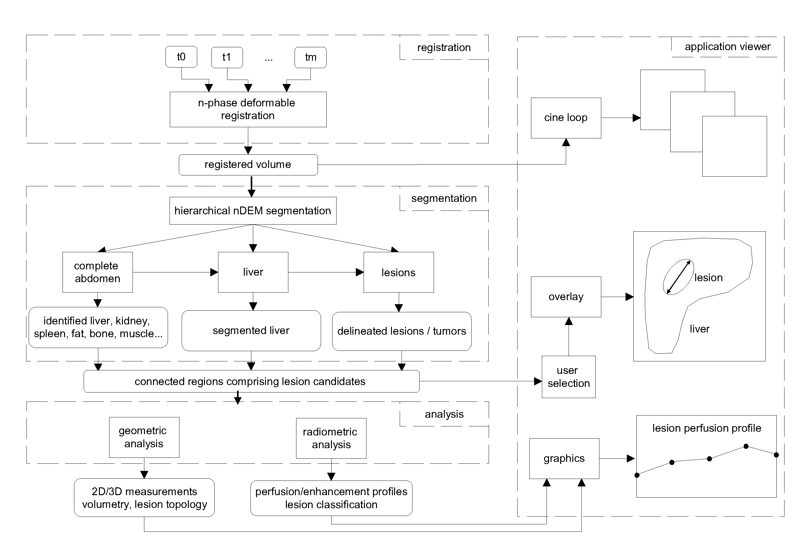
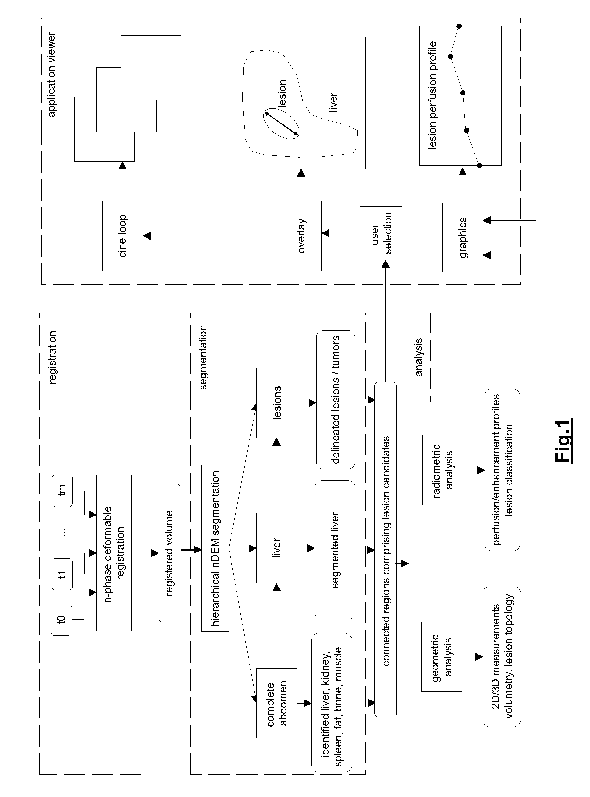
[0057]By way of example the application of the method of the invention to the liver and liver lesions is now described. The system and method of the invention can indeed advantageously be applied for analyzing multi-phase volumetric CT liver studies. The proposed approach greatly simplifies the state of the art diagnostic procedure for one or more longitudinal multi-phase studies by enabling the radiologist to synchronously assess a lesion over all phases and across longitudinal studies, by suggesting delineations obtained via automatic segmentation techniques, which simultaneously exploit the information in all the phases, and by providing accurate 3D measurements, such as the volume of the liver and the individual lesions and its changes in time, the longest diameter in 3D, the liver segment where the lesions are situated etc. FIG. 4 shows a different intra-study phases (without contrast enhancement: blanco at t=t0; with contrast enhancement: venous-portal at t=t2, venous at t=t3)...


PUM


Abstract

A method and system for acquiring information on lesions in dynamic 3D medical images of a body region and/or organ, having the steps: applying a registration technique to align a plurality of volumetric image data of the body region and/or organ, yielding multi-phase registered volumetric image data of the body region and/or organ; applying a hierarchical segmentation on the multi-phase registered volumetric image data of the body region and/or organ, the segmentation yielding a plurality of clusters of n-dimensional voxel vectors of the multi-phase aligned volumetric image; determining from the plurality of clusters a cluster or set of clusters delineating the body and/or organ; identifying the connected region(s) of voxel vectors belonging to the body region and/or organ; refining/filling the connected region(s) corresponding to the body region and/or organ; reapplying the segmentation step to the refined/filled connected region(s) corresponding to the body region and/or organ, to obtain a more accurate segmentation; and acquiring information on the presence of lesions in cluster of set of clusters of said plurality of clusters.

Description

FIELD OF THE INVENTION[0001]The present invention generally relates to the field of methods and devices for visualising medical image data of an examination volume in order to detect lesions.BACKGROUND OF THE INVENTION[0002]Nowadays, a standard way to assess certain pathologies and especially tumours in liver, is to use a four-phase contrast enhanced computer tomography (CT) protocol. This means that in one study four volumetric data sets are obtained: one before contrast injection and three after, when contrast agent is present at the hepatic artery, the portal vein and the hepatic vein. In general the contrast is the best at the portal-venous phase. In order to make a diagnosis the radiologist has to go through these four data sets and visually compare the lesions at corresponding positions by selecting the appropriate slices in each of the volumes individually. Once a tumour is discerned, its diameter will be measured according to the RECIST (Response Evaluation Criteria In Solid...

Claims


Application Information

Patent Timeline
IPC IPC(8): G06K9/00
CPCG06T3/0081G06T7/0012G06T7/401G06T2200/24G06T2207/30056G06T2207/20036G06T2207/20096G06T2207/30004G06T2207/10072G06T7/41
Inventor MARKOVA, ANETADEKLERCK, RUDIDEMEY, JOHANCLERINX, PETERPOLE, IANDEWAELE, PIET
Owner IBBT
Why Eureka
Try Eureka