

Anatomy-defined automated cpr generation
anatomy-defined, automated technology, applied in the direction of 3d-image rendering, instruments, computing, etc., can solve the problem that it is not easy to adapt to visualizing other objects such as the human heart or brain
- Summary
- Abstract
- Description
- Claims
- Application Information
AI Technical Summary
Benefits of technology
Problems solved by technology
Method used
Image

Examples
Embodiment Construction
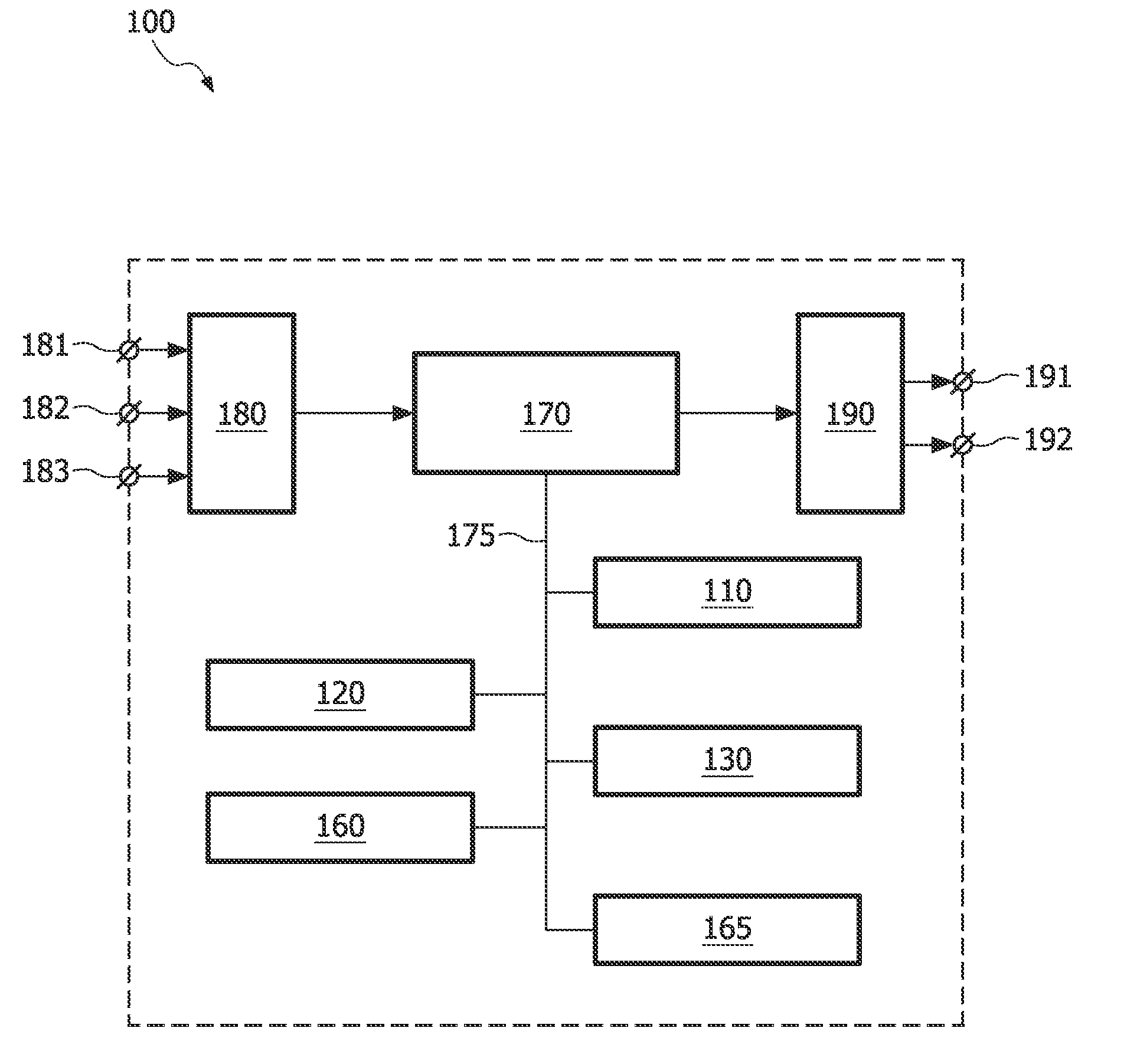
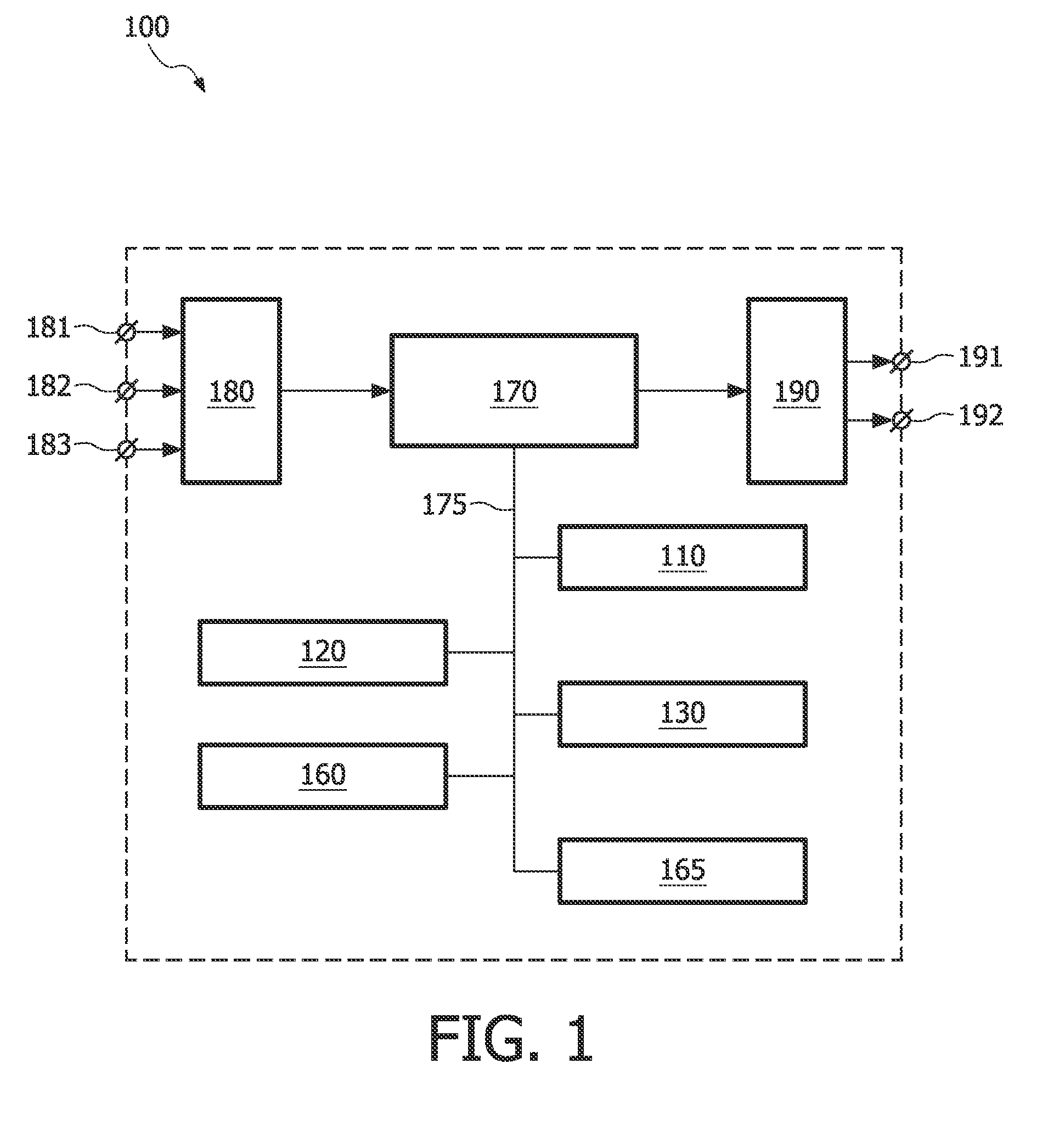
[0040]FIG. 1 schematically shows a block diagram of an exemplary embodiment of the system 100 for visualizing an object in image data using a first cross-section surface coupled to a model of the object, the system comprising:
[0041]a model unit 110 for adapting the model to the object in the image data;
[0042]a surface unit 120 for adapting the first cross-section surface to the adapted model on the basis of the coupling between the first cross-section surface and the model; and
[0043]a visualization unit 130 for computing an image from the image data on the basis of the adapted first cross-section surface.
[0044]The exemplary embodiment of the system 100 further comprises the following units:
[0045]a control unit 160 for controlling the workflow in the system 100;
[0046]a user interface 165 for communicating with a user of the system 100; and
[0047]a memory unit 170 for storing data.
[0048]In an embodiment of the system 100, there are three input connectors 181, 182 and 183 for the incomi...
PUM
 Abstract
Description
Claims
Application Information