Quantification method of the feature of a tumor and an imaging method of the same

Active Publication Date: 2011-07-28
AMCAD BIOMED CORP
View PDF21 Cites 24 Cited by
  • Summary
  • Abstract
  • Description
  • Claims
  • Application Information

AI Technical Summary

Benefits of technology

[0030]As a result, with the quantification method of the margin feature of a tumor, the imaging method of the margin feature of a tumor, the quantification method of the cysts feature of a tumor, the imaging method of the cysts feature of a tumor, the quantification method of the calcifications feature of a tumor, the imaging method of the calcifications feature of a tumor, the quantification method of the echoic feature of a tumor, the quantification method of the heterogenesis feature of a tumor, and the imaging method of the heterog

Problems solved by technology

However, the medical use ultrasonic imaging system of the present time can only allow the doctor to input the tumor contour identified by his naked eyes to the ultrasonic image of the tumor, with the help of a handwriting input device.
But, the unreliability of the diagnosis of the nature of the tumor is significant just due to this tumor contour identifying process, for the reasons below:
Therefore, mistakes on diagnosis of the nature of a tumor exist from time to time, decreasing the reliability of the diagnosis process of the nature of a tumor by the m

Method used


Image

  • Quantification method of the feature of a tumor and an imaging method of the same
  • Quantification method of the feature of a tumor and an imaging method of the same
  • Quantification method of the feature of a tumor and an imaging method of the same

Examples


first embodiment

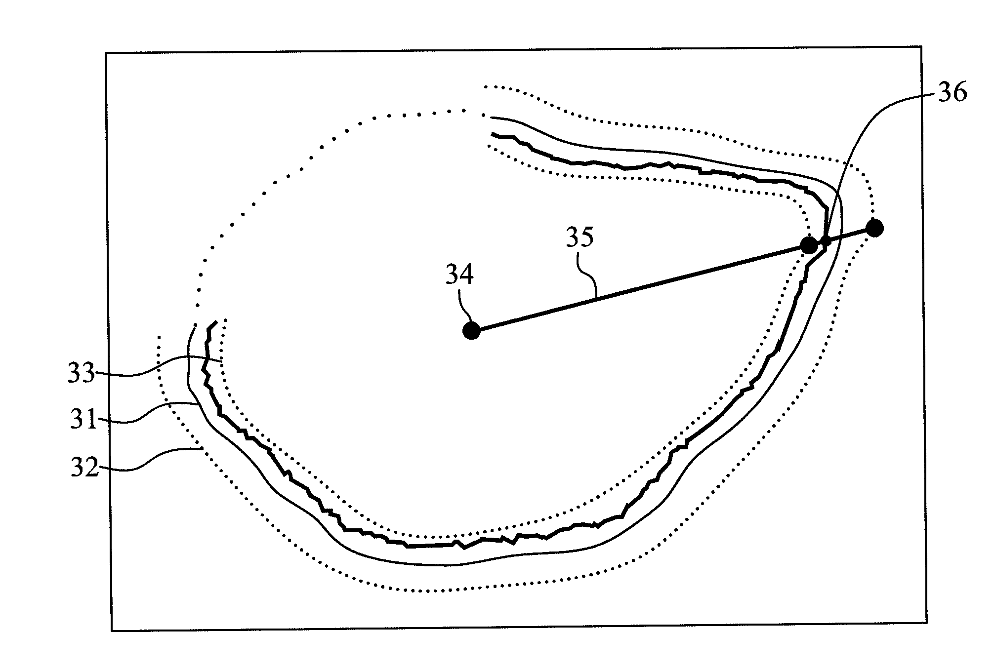
[0067]Besides, as shown in FIG. 3B, which is a flowchart of the quantification method of the margin feature of a tumor according to the present invention, comprising the steps of:

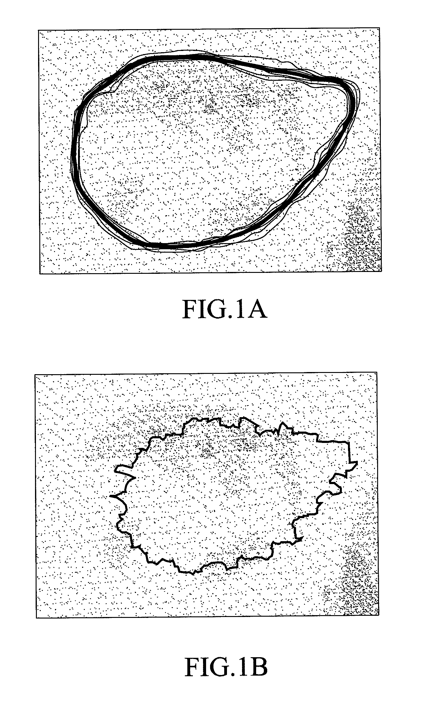
[0068](A) retrieving a tumor contour and a tumor contour annular region from the gray scale image, wherein the tumor contour is in the tumor contour annular region;

[0069](B) displaying the tumor contour over the gray scale image for defining a tumor inner region and a tumor external region on the gray scale image;

[0070](C) retrieving a center of gravity of the tumor contour annular region, defining a section line extending outwardly from the center of gravity and penetrating the tumor contour annular region, and providing a measured line segment being on the section line and in the tumor contour annular region;

[0071](D) calculating the moving variance of the gray scale of each of the plurality pixel points on the measured line segment; and

[0072](E) quantifying the margin feature of the tumor on the section ...

second embodiment

[0093]As shown in FIG. 4A, which is a flowchart of the imaging method of the margin feature of a tumor according to the present invention, comprising the steps of:

[0094](A) retrieving a tumor contour and a tumor contour annular region from the gray scale image, wherein the tumor contour is in the tumor contour annular region;

[0095](B) displaying the tumor contour over the gray scale image for defining a tumor inner region and a tumor external region on the gray scale image;

[0096](C) retrieving a center of gravity of the tumor contour annular region, defining a section line extending outwardly from the center of gravity and penetrating the tumor contour annular region, and providing a measured line segment being on the section line and in the tumor contour annular region;

[0097](D) calculating the moving variance of the gray scale of each of the plurality pixel points on the measured line segment; and

[0098](E) defining a margin imaging upper limit and a margin imaging lower limit base...

third embodiment

[0112]As shown in FIG. 6B, which is a flowchart of the quantification method of the cysts feature of a tumor according to the present invention, comprising the steps of:

[0113](A) retrieving a tumor contour and a tumor contour annular region from the gray scale image, wherein the tumor contour is in the tumor contour annular region;

[0114](B) displaying the tumor contour over the gray scale image for defining a tumor inner region and a tumor external region on the gray scale image;

[0115](C) calculating both the minimum value and the standard deviation of the gradient value of the gray scale of the plurality pixel points in the tumor inner region; and

[0116](D) quantifying the cysts feature of the tumor in the tumor inner region, based on both the minimum value and the standard deviation of the gradient value of the gray scale of the plurality pixel points in the tumor inner region.

[0117]However, since the tumor contour and the tumor contour annular region retrieved from the gray scale ...


PUM


Abstract

A quantification method and an imaging method are disclosed, capable of quantifying the margin feature, the cysts feature, the calcifications feature, the echoic feature and the heterogenesis feature of a tumor, and capable of imaging the margin feature, the cysts feature, the calcifications feature and the heterogenesis feature of a tumor. The quantification method and the imaging method calculate the moving variance of the gray scale of each of the pixel points based on the gradient value of the gray scale of these pixel points. Then, depending on the purpose of the quantification method or the imaging method, the maximum value, the minimum value, the mean value, and the standard deviation of the moving variance of the gray scale of these pixel points are calculated, respectively. At final, with the definition of the threshold value and the imaging rule, the above features of the tumor are quantified or imaged.

Description

BACKGROUND OF THE INVENTION[0001]1. Field of the Invention[0002]The present invention relates to a quantification method and an imaging method, and more particularly, to the quantification method capable of quantifying the margin feature, the cysts feature, the calcifications feature, the echoic feature and the heterogenesis feature of a tumor, and to the imaging method capable of imaging the margin feature, the cysts feature, the calcifications feature and the heterogenesis feature of a tumor.[0003]2. Description of Related Art[0004]In recent years, since the resolution of the image and the digitized process of the image of the medical use ultrasonic imaging technology have both increased significantly, the application filed of the medical use ultrasonic imaging technology is not limited in monitoring the growth of a baby anymore. The medical use ultrasonic imaging technology is gradually used in the diagnosis of the nature of many kinds of tumor, such as the thyroid tumor. Moreove...

Claims


Application Information

Patent Timeline
IPC IPC(8): G09G5/02G09G5/00
CPCG06T7/0012G06T7/0083G06T2207/30096G06T7/401G06T2207/10132G06T7/0087G06T7/12G06T7/143G06T7/41
Inventor CHANG, KING JENCHEN, WEN HWACHEN, ARGONCHEN, CHIUNG NEINHO, MING CHIHTAI, HAO CHIHWU, MING HSUNTSAI, PO WEILIU, CHUNG WEIWU, HSIN-JUNG
Owner AMCAD BIOMED CORP
Why Eureka
Try Eureka