Natural Polymer-Based Porous Orthopedic Fixation Screw for Bone Repair and Regeneration
- Summary
- Abstract
- Description
- Claims
- Application Information
AI Technical Summary
Benefits of technology
Problems solved by technology
Method used
Image


Examples
example 1
Microsphere Fabrication
[0061]Cellulose acetate (CA) or ethyl cellulose microspheres (EC) are fabricated using an oil-in-water emulsion / solvent evaporation method. In brief, either CA or EC is dissolved in a binary solvent composition of methylene chloride:acetone (9:1) at 20% (w / v). The resulting polymer solution is slowly poured into a 1% (w / v) polyvinyl alcohol aqueous solution stirring at 250 rpm. The solvent is allowed to evaporate overnight at room temperature under constant stirring. The microspheres are collected by vacuum filtration and washed with distilled water. Microspheres are sieved and separated into different sizes based on their diameter for scaffold fabrication. Three different diameters namely >1180, 1180-850, and 850-600 μm were chosen for use in bone grafts. While particles in the diameter range of 50-100 100-150, 200-250, 250-400 μm were chosen for fabricating fixation devices.
example 2
Scaffold Fabrication Using Solvent / Non-Solvent Sintering
[0062]It is necessary to identify a proper solvent / non-solvent composition for each polymer at which only the microsphere surface turns rubbery to facilitate bonding with the adjacent microspheres. After several trials a solvent / non-solvent composition of 3:1 ratio of acetone:water was found to be suitable for ethylcellulose (EC) microsphere sintering while 8:2 ratio of acetonitrile:water for cellulose acetate (CA). The sieved microspheres were mixed with sintering solvent and the mixture was vortexed for five seconds. The resulting slurry was placed in a cylindrical Teflon mold with a 5 mm diameter and 10 mm height. The solvent / non-solvent mixture was allowed to evaporate in a fume hood for 30 minutes followed by vacuum-drying for an additional 24 hours. Scaffolds of 8 mm diameter and 2 mm thickness were also fabricated for in vitro cell studies. In contrast control poly(lactide-co-glycolide) (PLAGA) sintered microsphere matri...
example 3
3-D Sintered Microsphere Characterization Morphology
[0065]3-D Composite microsphere scaffold morphology is characterized by SEM. Cylindrical scaffolds (n=3) are coated with gold using a Hummer V sputtering system (Technics, Baltimore, Md.) for 5 min. Samples are visualized on a JSM 6400 (JOEL, Boston, Mass.) at 15-20 keV and a working distance of 39-48 cm.
[0066]Scanning electron microscopy (SEM) is used to characterize the morphology of the individual microspheres and the corresponding scaffolds.
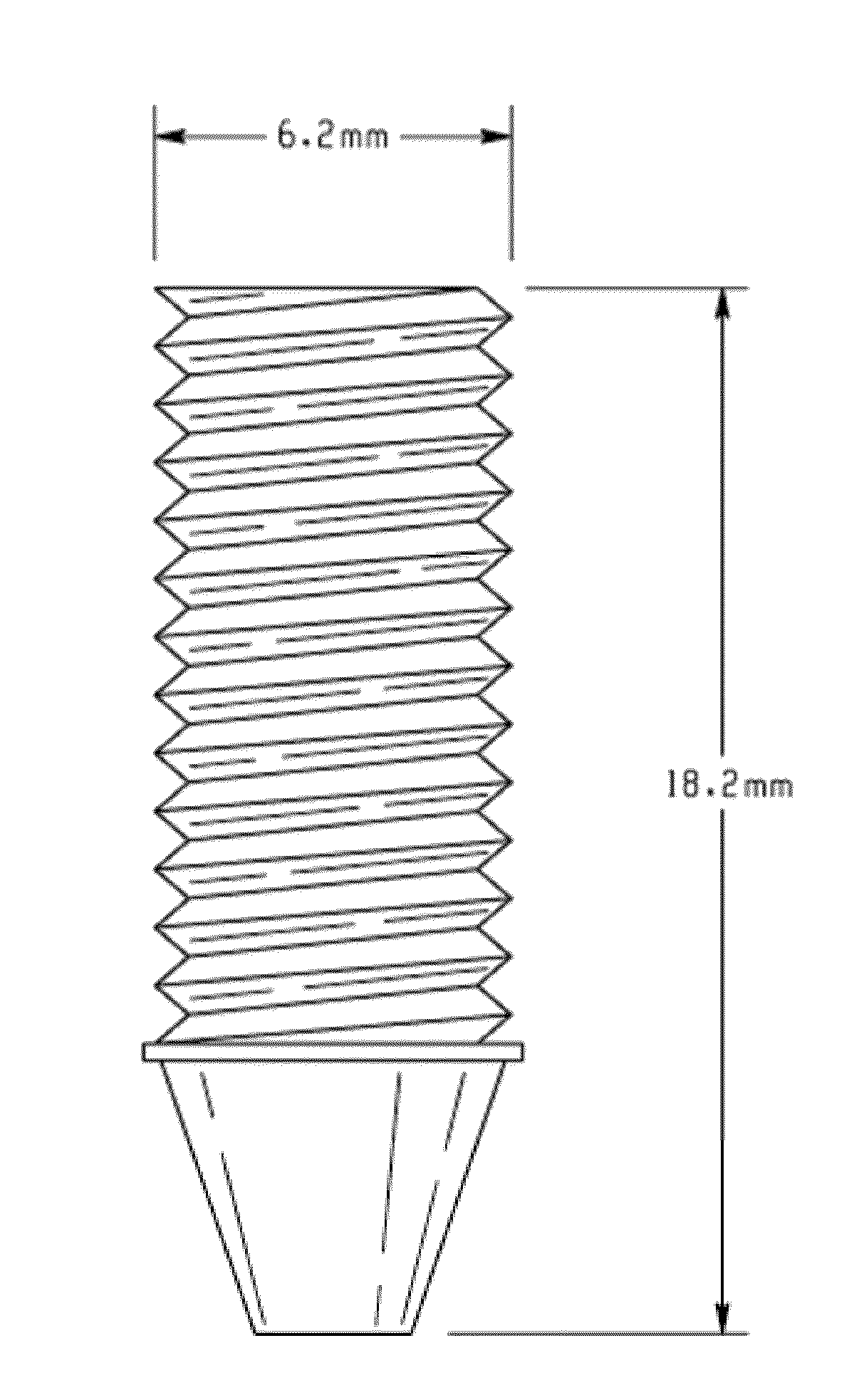
[0067]TABLE I provides a summary of the mechanical properties of cylindrical and interference screw structures fabricated from CA and CA-HA under compression, bending and torsional modes.
MolecularCompressiveCompressiveweightModulusStrengthCellulose Structure(Mn)(MPa)(MPa)Cylinder30,000257 ± 2216 ± 4Cylinder50,000366 ± 2133 ± 8Screw30,000415 ± 1317 ± 3Screw50,000422 ± 17 28 ± 10CA-15 wt % HA Screw30,000389 ± 1614 ± 3CA-15 wt % HA Screw50,000395 ± 2017 ± 5PLGA Cylinder190,000155 ± 30 4 ± 1PLGA...
PUM
| Property | Measurement | Unit |
|---|---|---|
| Temperature | aaaaa | aaaaa |
| Temperature | aaaaa | aaaaa |
| Percent by mass | aaaaa | aaaaa |
Abstract
Description
Claims
Application Information