Method for identifying antigen-specific regulatory t cells

a technology of antigen-specific regulatory cells and t cells, which is applied in the field of methods for identifying and isolating antigen-specific regulatory t cells, can solve the problems of unable to isolate viable treg populations for functional studies or ex vivo expansion as a prelude to therapeutic administration, and the accuracy of identifying viable treg using cd4, cd25 and foxp3 markers is problemati

Inactive Publication Date: 2011-09-22
ST VINCENTS HOSPITAL SYDNEY
View PDF4 Cites 8 Cited by
  • Summary
  • Abstract
  • Description
  • Claims
  • Application Information

AI Technical Summary

Problems solved by technology

However, there are a number of issues that are currently impeding Treg therapy.
First, accurate identification of viable Treg using the CD4, CD25 and Foxp3 markers is problematical.
Further, detection of Treg using Foxp3 antibodies requires fixation and permeabilisation of the cells, so the technique cannot be used to isolate viable Treg populations for functional studies or ex vivo expansion as a prelude to therapeutic administration.
Secondly, Treg are present in low numbers in the circulation.
However, methods of detecting the antigen specificity of Treg have not previously been described.

Method used


Image

  • Method for identifying antigen-specific regulatory t cells
  • Method for identifying antigen-specific regulatory t cells
  • Method for identifying antigen-specific regulatory t cells

Examples


example 1

Antigen Stimulation and Cell Identification and Isolation

[0133]Materials and Methods

[0134]Reagents

[0135]The antigens used were cytomegalovirus (CMV) lysate (22), mycobacterial antigen lysates (Mycobacterial tuberculosis (MTB) or Mycobacterial Avium intracellular complex (MAI), CSL, Melbourne, Australia), CMV-peptide (P1) (23, 24). Staphylococcal enteroantigen B (SEB) (Sigma-Aldrich Co., St Louis, Mo., United States of America) was used as a mitogen.

[0136]The monoclonal antibodies (mAbs) used were anti-CD3-PerCP-Cy5.5, anti-CD4-PE-Cy7, anti-CD45RO-FITC, anti-CD25-APC, anti-CD134-FITC, anti-CD134-PE and anti-Foxp3 (clone 259D) (all from Becton-Dickinson, San Jose, Calif., United States of America); anti-CD45RO-ECD (Beckman Coulter, Hialeah, Fla., United States of America); CD127-PE (Immunotech, Marseille, France); anti-CD127-Pacific Blue and anti-Foxp3-APC (clone PCH101) (eBiosciences, San Diego, Calif., United States of America); and CD39-PE (Serotec, Oxford, UK). All antibodies were...

example 2

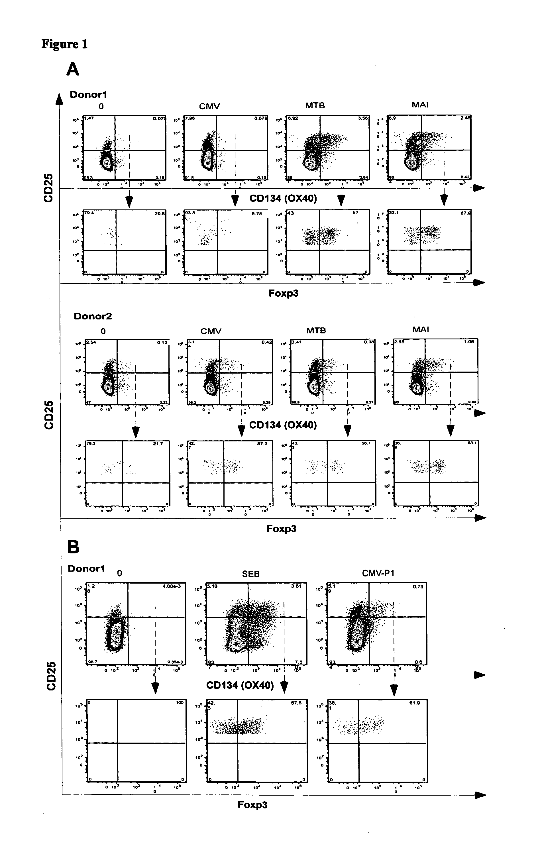
Identification of CD4+CD25+CD134+Foxp3+ Antigen-specific Treg from Whole Blood

[0141]Foxp3 is an accepted marker of Treg. It was investigated whether antigen-specific CD4+ Treg could be identified in whole blood by detecting the co-expression of CD4, CD134, CD25 and Foxp3 following in vitro stimulation with antigen

[0142]Materials and Methods

[0143]Whole blood from two healthy controls (donor 1 and donor 2), who were known to have active immune responses to MAI, MTB or CMV was stimulated in vitro with no antigen, CMV, MTB or MAI lysates as described in Example 1. In a second experiment, whole blood from donor 1 was stimulated with no antigen, CMV-P1 antigen or SEB mitogen (as a positive control) for 24 to 40 hours as described in Example 1.

[0144]The cells were then stained with the combination of mAbs consisting of anti-CD3, anti-CD4, anti-CD25, anti-CD134 and anti-Foxp3 antibodies. Cell staining was analysed on a three-laser LSR II flow cytometer (Becton-Dickinson). Antigen-specific T...

example 3

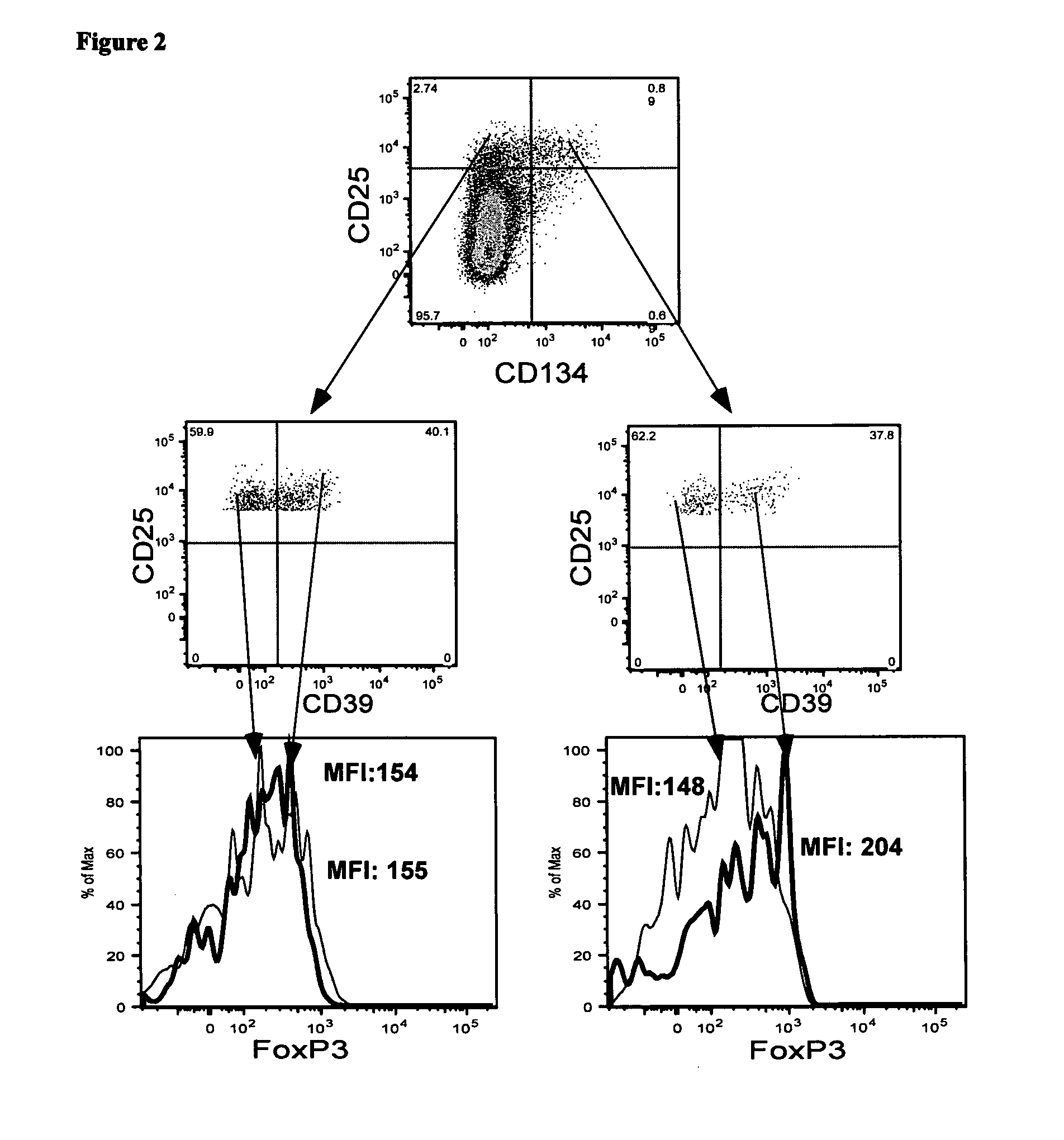
Isolation of CD4+CD25+CD39+CD134+ Viable Antigen-Specific Treg from Whole Blood

[0151]Detection of Foxp3 requires cell permeabilisation, which is problematical when it is desirable to have live cells following cell staining. CD39 has recently been reported to be a useful alternative marker for detecting Treg. In this example, the present applicant investigated whether antigen-specific CD4+Treg could be identified in whole blood following in vitro stimulation with antigen by detecting the co-expression of CD4, CD25, CD134 and CD39.

[0152]Materials and Methods

[0153]Whole blood from donor 1 was stimulated with no antigen, CMV-P1 antigen or SEB mitogen (as a positive control) for 24 hr as described in Examples 1 and 2. The cells were then stained with the monoclonal antibodies consisting of anti-CD3, anti-CD4, anti-CD39, anti-CD25, anti-CD134 and anti-Foxp3 as described in Examples 1 and 2. CD3+CD4+ cells were gated.

[0154]Results

[0155]The plots shown in FIG. 2 demonstrate that approximate...


PUM

PropertyMeasurementUnit
timeaaaaaaaaaa
timeaaaaaaaaaa
timeaaaaaaaaaa

Abstract

A method of identifying an antigen-specific regulatory T cell (Treg) from a subject is discussed wherein the method comprises quantitatively or qualitatively detecting co-expression of each of cell markers CD4, CD25 and CD134, or alternatively, N each of cell markers CD8, CD25 and CD137, as well as one or more cell markers selected from the group of Treg cell markers consisting of CD39, CD73, CD127, CTLA-4 and Foxp3 on a cell in a suitable lymphocyte-containing sample from the subject in response to exposure to a target antigen. Also discussed are methods of isolating and expanding the identified antigen-specific Treg population, which may permit antigen-specific Treg cell therapy.

Description

INCORPORATION BY REFERENCE[0001]The following co-pending patent applications are referred to in the following description:[0002]PCT / AU2006 / 001080 (WO 2007 / 014420) titled “METHOD FOR IDENTIFYING REGULATORY T CELLS”; and[0003]PCT / AU2007 / 000342 (WO 2007 / 106939) titled “A METHOD FOR DETECTING ANTIGEN SPECIFIC OR MITOGEN ACTIVATED T CELLS”.[0004]The entire content of both of these applications is hereby incorporated by reference.FIELD OF THE INVENTION[0005]The present invention relates to methods for identifying and isolating antigen-specific regulatory T cells.PRIORITY DOCUMENTS[0006]The present application claims priority from:[0007]Australian Provisional Patent Application No. 2007905154 entitled “A Method” and filed on 20 Sep. 2007; and Australian Provisional Patent Application No. 2007905292 entitled “A Method” and filed on 27 Sep. 2007. The entire content of each of these applications is hereby incorporated by reference.BACKGROUND OF THE INVENTION[0008]T cells are a type of lymphoc...

Claims


Application Information

Patent Timeline
Patent Type & Authority Applications(United States)
IPC IPC(8): A61K35/14C12Q1/02C12N5/0783A61P37/04
CPCG01N33/505A61P37/04
Inventor KELLEHER, ANTHONY DOMINICZAUNDERS, JOHN JAMESSEDDIKI, NABILA
Owner ST VINCENTS HOSPITAL SYDNEY
Why Eureka
Try Eureka