Method for tissue characterization based on beta radiation and coincident Cherenkov radiation of a radiotracer

a radiotracer and tissue characterization technology, applied in the field of tissue characterization, can solve the problems of significant cost, difficult to distinguish the outer periphery from the healthy tissue, and significant time consumption

Inactive Publication Date: 2011-10-13
THE BOARD OF TRUSTEES OF THE LELAND STANFORD JUNIOR UNIV
View PDF1 Cites 10 Cited by
  • Summary
  • Abstract
  • Description
  • Claims
  • Application Information

AI Technical Summary

Problems solved by technology

Although effective in the bulk tumor, the outer periphery is difficult to discriminate from the healthy tissue, due to the microscopic involvement.
This process is laborious, and involves a significant amount of time, which comes at significant cost.
Furthermore, this process involves a difficult trade-off between resecting too little, at a risk of not extracting all the tumor tissue, or resecting too much, causing unnecessary harm to the patient.

Method used


Image

  • Method for tissue characterization based on beta radiation and coincident Cherenkov radiation of a radiotracer
  • Method for tissue characterization based on beta radiation and coincident Cherenkov radiation of a radiotracer
  • Method for tissue characterization based on beta radiation and coincident Cherenkov radiation of a radiotracer

Examples


Embodiment Construction

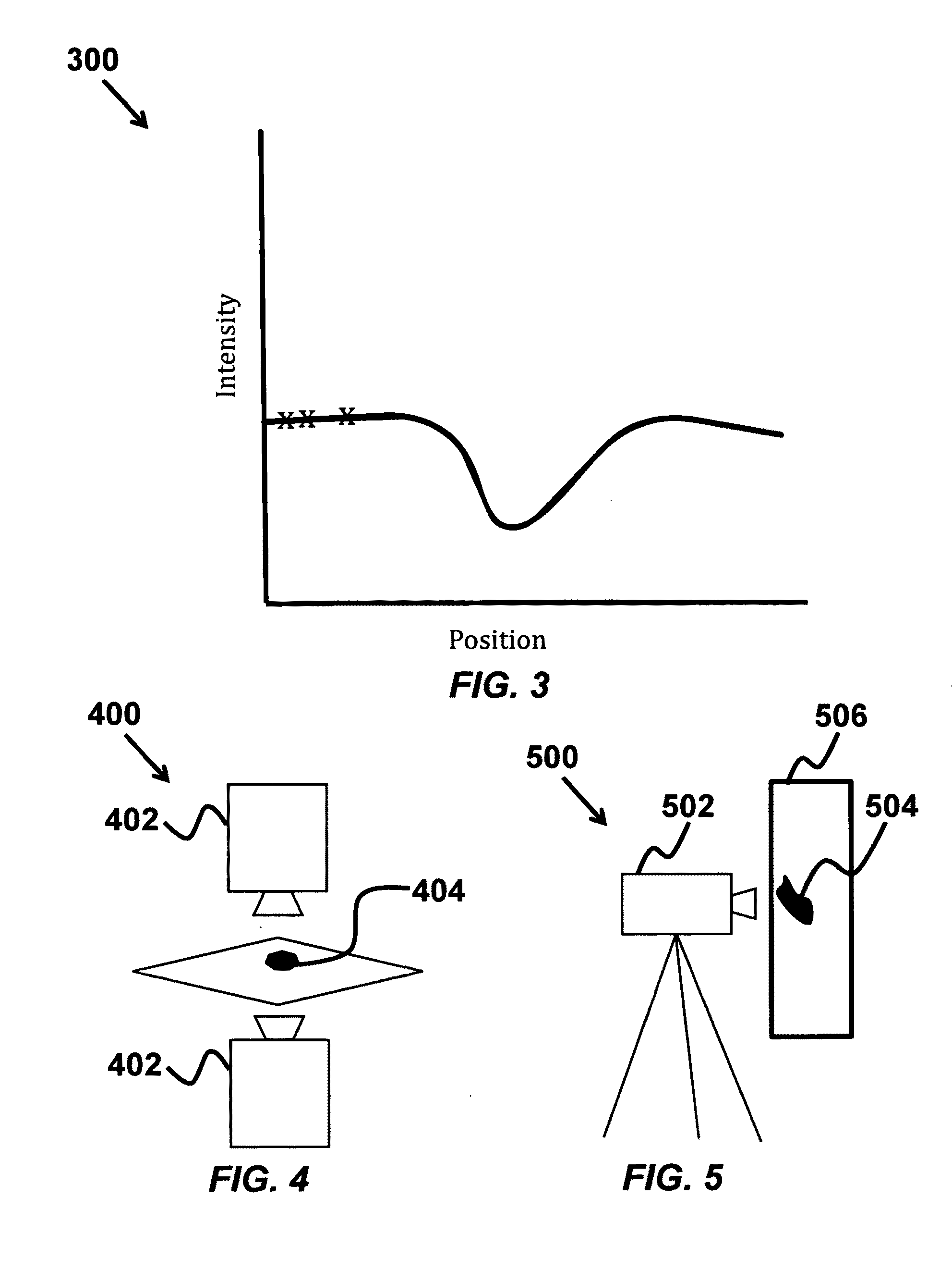
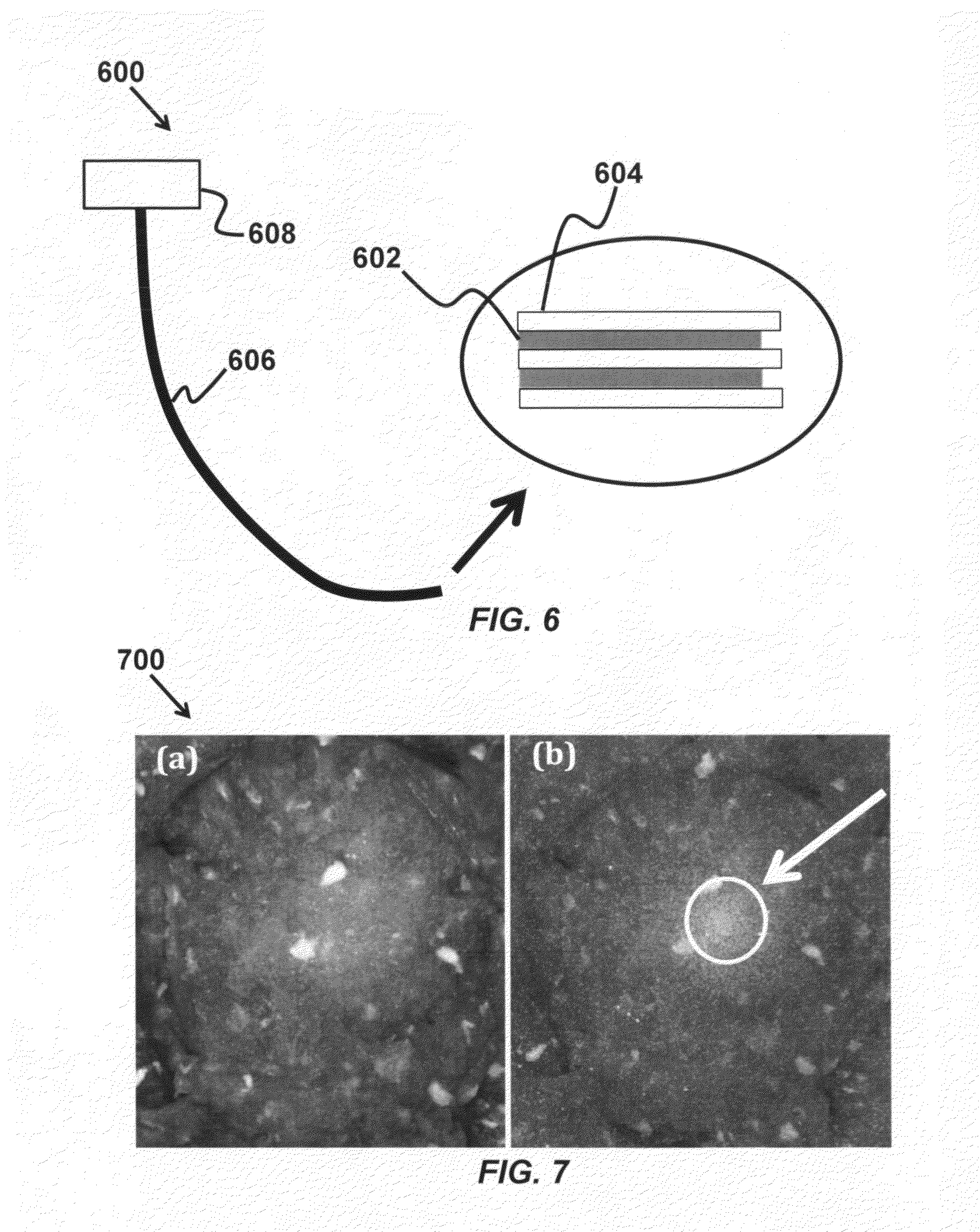
[0018]This invention is a method to image excised or superficial tissue with high resolution, high sensitivity, and high specificity, which combines the micrometer-resolution of optical imaging with the specificity of disease-specific radiotracers. This is accomplished by imaging the emitted beta and / or Cherenkov radiation, which provide depth-selective imaging, although this technique is not restricted to depth-selective imaging. The optical tissue characterization via Cherenkov radiation is used to determine disease status by the high resolution imaging of targeted radiotracers in tissue samples. This characterization is used for disease diagnosis, both in vivo and in vitro, which allows on-site delineation of surgical margins during disease resection.

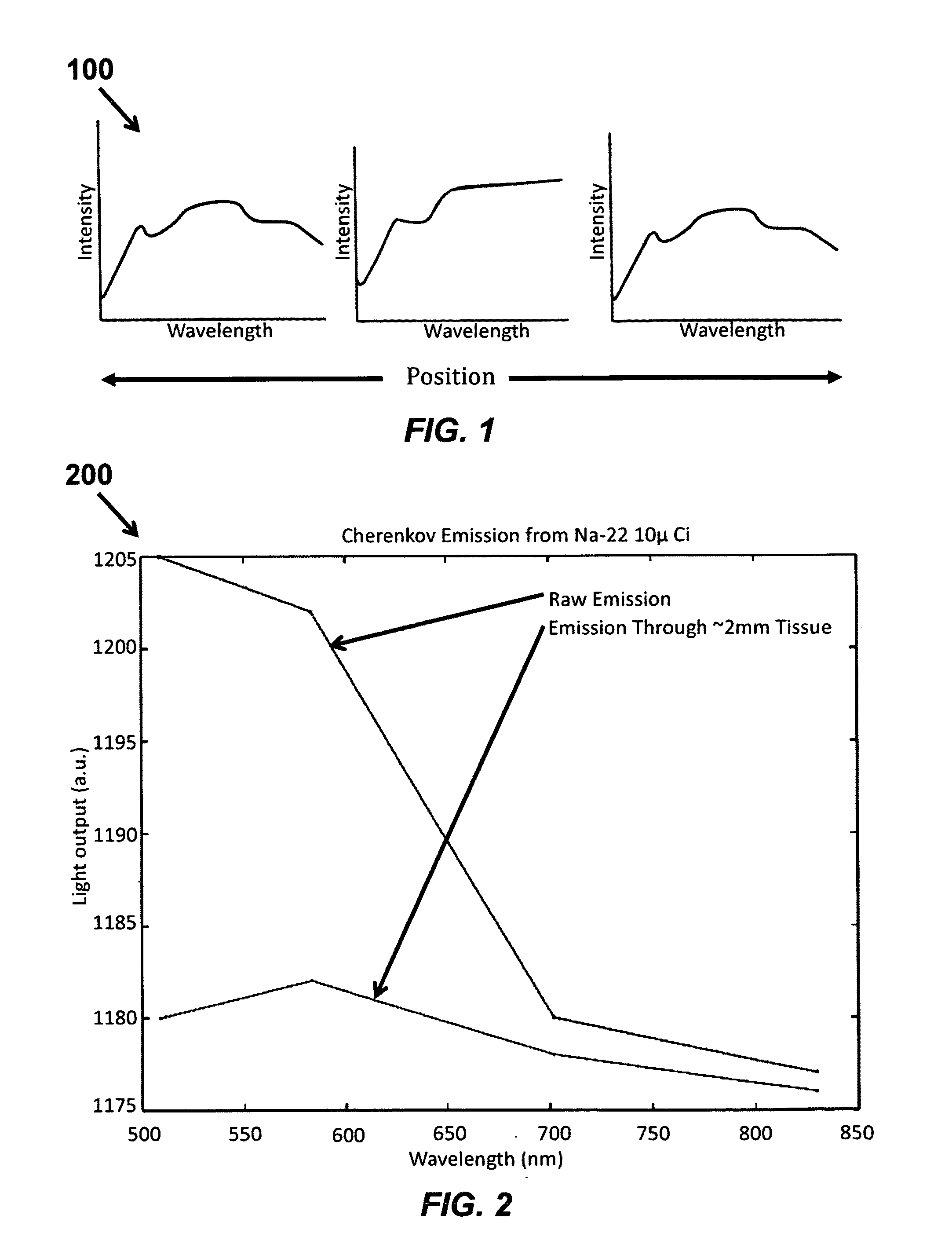
[0019]Radiotracers that are beta emitters will emit beta particles as they decay. These charged particles will radiate light if they exceed the apparent speed of light in a medium. Both the emitted light, which has a dominant blue co...


PUM

PropertyMeasurementUnit
depthaaaaaaaaaa
Cherenkov radiationaaaaaaaaaa
concentrationaaaaaaaaaa

Abstract

A method of characterizing a tissue sample is provided that includes injecting a tissue sample with radiotracers, where the radiotracers include beta-emitter radio tracers, the beta-emitter radio tracers emit beta particles according to a decay of the beta-emitter radio tracers, and measuring the beta particles or Cherenkov radiation from the beta particles in the tissue sample, and determining a condition of the radio tracers in the tissue sample according to the measured beta particles or the measured Cherenkov radiation, where the determined condition includes a depth and/or a concentration of the radiotracers in the tissue sample.

Description

CROSS-REFERENCE TO RELATED APPLICATIONS[0001]This application claims priority from U.S. Provisional Patent Application 61 / 342,321 filed Apr. 12, 2010, which is incorporated herein by reference.STATEMENT OF GOVERNMENT SPONSORED SUPPORT[0002]This invention was made with Government support under contract CA133474 awarded by National Institutes of Health, and under contract 0854492 awarded by National Science Foundation. The Government has certain rights in this invention.FIELD OF THE INVENTION[0003]The invention relates to tissue characterization. More specifically, the invention relates to methods for tissue characterization based on beta and Cherenkov radiation of molecular-specific radiotracers.BACKGROUND OF THE INVENTION[0004]Surgical excision of diseased tissue, such as malignant tumors, requires the surgeon to identify abnormal tissue based on physical appearance. Although effective in the bulk tumor, the outer periphery is difficult to discriminate from the healthy tissue, due t...

Claims


Application Information

Patent Timeline
Patent Type & Authority Applications(United States)
IPC IPC(8): A61K51/00C12Q1/02
CPCA61K51/04
Inventor CARPENTER, COLIN MXING, LEISUN, CONROY GHIN CHEEPRATX, GUILLEM
Owner THE BOARD OF TRUSTEES OF THE LELAND STANFORD JUNIOR UNIV
Why Eureka
Try Eureka