System and method for imaging myocardial infarction

a technology of myocardial infarction and functional analysis, applied in the field of medical diagnostic and screening systems and methods, can solve the problems of unsatisfactory none of these strategies have produced a solution to the high incidence of death due to undetected cad, and the need for improved cardiac imaging and functional analysis systems and methods remains a tremendous unmet need, so as to reduce image distortion, minimize valve phase map distortion, and enhance valve plane delin

Inactive Publication Date: 2008-08-05
BISHOP HARRY A
View PDF2 Cites 11 Cited by
  • Summary
  • Abstract
  • Description
  • Claims
  • Application Information

AI Technical Summary

Benefits of technology

[0009]An advantage of the invention is that it utilizes first pass rather than equilibrium pool planar angiography. This results in greatly reduced image distortion due to such factors as respiratory motion and crosstalk. The small changes in myocardial motion due to infarction otherwise can be completely obscured by image distortions inherent in standard equilibrium (MUGA) phase measurements.
[0010]Another advantage of the invention is the timing precision (temporal resolution) of the cyclic change in blood tracer is accurate to 0.001 second. Also, data is acquired in list mode rather than in frame mode.
[0011]Another advantage of the invention is that it uses a 30 degree right anterior oblique view of the heart which is optimal for visualizing the maximum extent of myocardial wall in profile and the heart valves are nearly perpendicular to the imaging plane. This minimizes the valve distortion of the phase maps of the ventricular blood pool and enhances valve plane delineation by functional imaging.
[0

Problems solved by technology

While cardiac imaging and functional analysis is the largest single nuclear medical imaging application, there remains a tremendous unmet need for improved cardiac imaging and functional analysis systems and methods.
None of these strategies h

Method used


Image

  • System and method for imaging myocardial infarction
  • System and method for imaging myocardial infarction
  • System and method for imaging myocardial infarction

Examples


Embodiment Construction

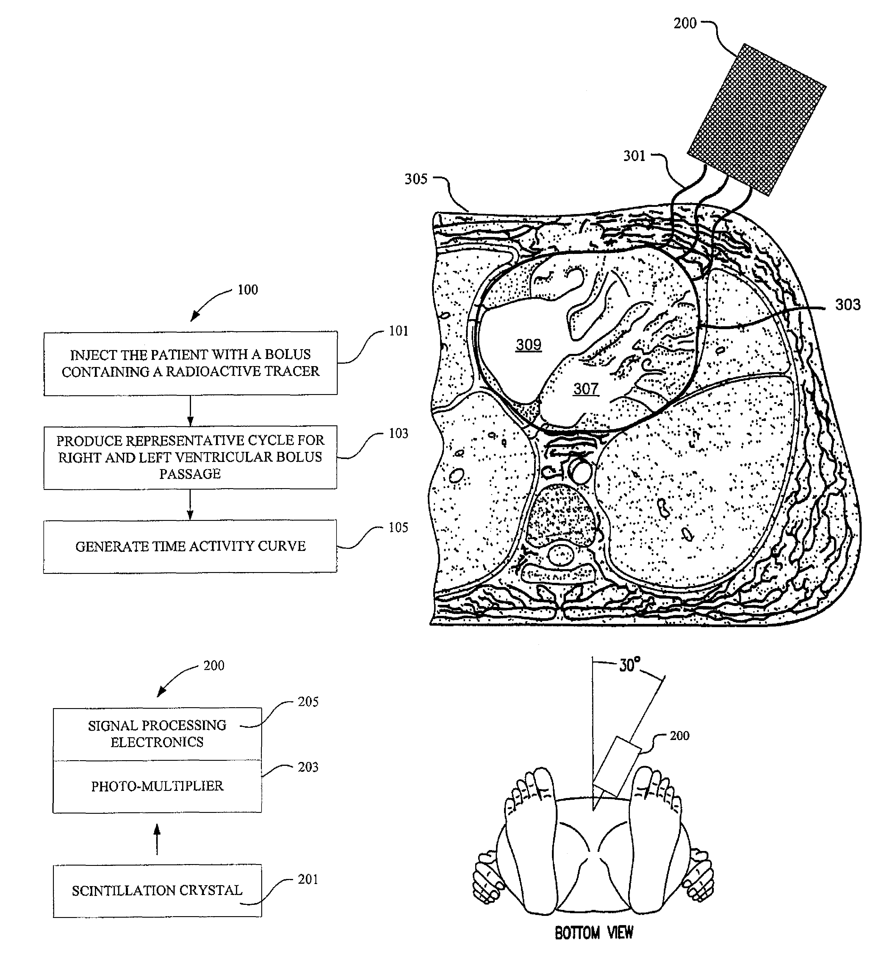
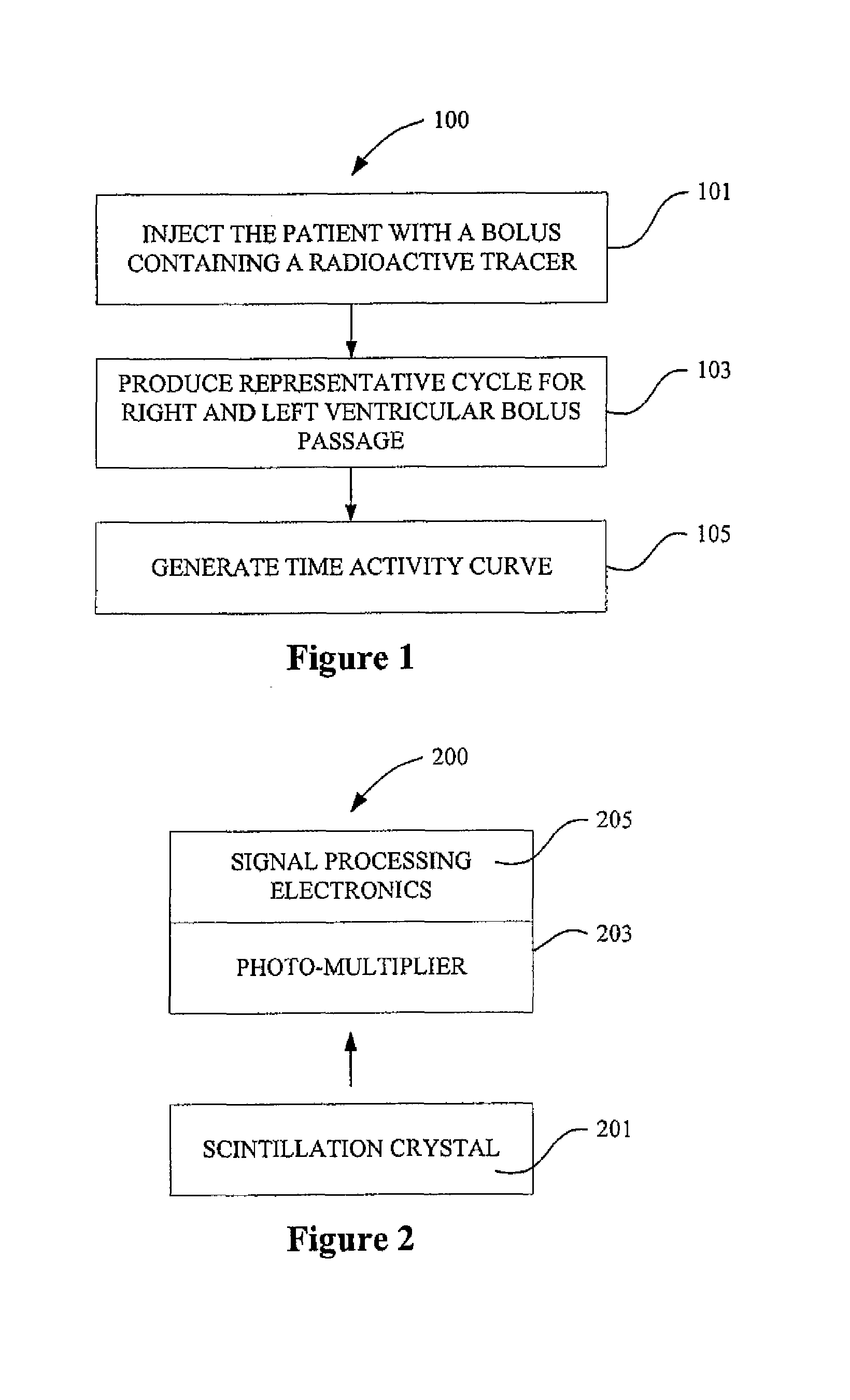
[0027]FIG. 1 provides a flow chart illustrating a method 100 of imaging a myocaridal infarction in a patient in accordance with one aspect of the present invention. The method involves action 101, injecting a patient with a radioactive bolus, action 103, producing a representative cycle for right ventricular bolus passage and a representative cycle for left ventricular passage, and action 105, generating a time activity curve based on activity in each segment of the respective representative cycles.

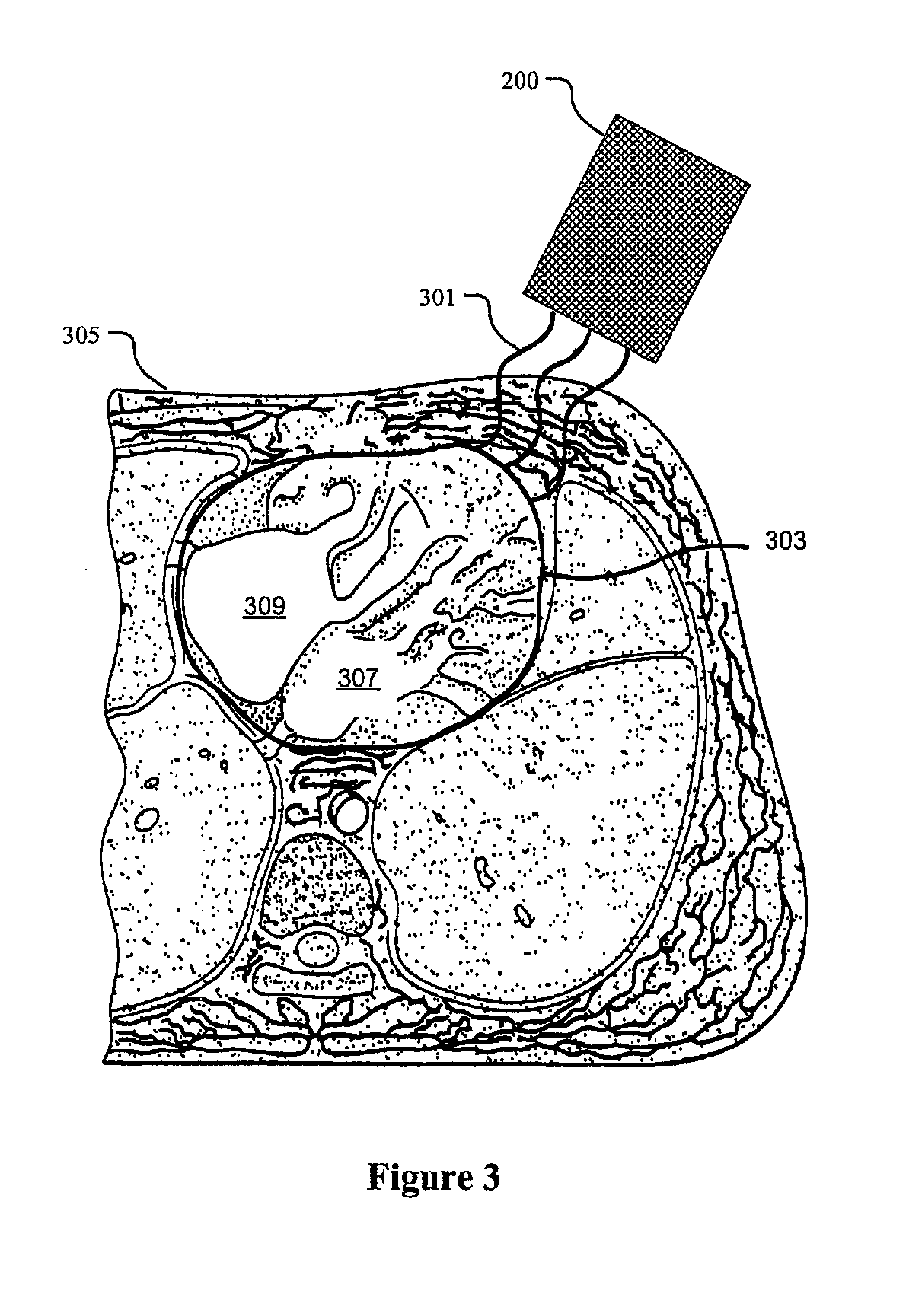
[0028]Acute myocardial infarction characteristically presents with an acute onset of severe chest pain at rest. The present method 100 is used to delineate the infarction-produced focal defect in myocardial contractility by producing a phase map of the ventricular blood flow through a patient's heart that shows the location and extent of the myocardial damage to the heart. The method 100 thus can inform a health care provider whether a patient's chest pain is due to a myocardial infarctio...


PUM


Abstract

One aspect of the present invention relates to imaging a patient to determine whether he or she has suffered a myocardial infarction. According to this method, a patient is injected with a bolus having a radioactive tracer. A representative cycle is produced for both the right ventricular passage of the bolus and the left ventricular passage of the bolus based on planar coordinates over time of scintillation events of the tracer. A time activity curve based on activity in each segment of the respective representative cycles is generated. When a segment of heart muscle is damaged its contraction ceases or lags behind the normal surrounding myocardium resulting in a phase shift in the adjacent blood pool.

Description

CROSS REFERENCE TO RELATED APPLICATIONS[0001]This invention claims priority to U.S. Patent Application No. 60 / 543,438 filed Feb. 9, 2004.FIELD OF INVENTION[0002]The present invention relates to medical diagnostic and screening systems and methods. In particular, the present invention relates to non-invasive cardiac imaging and functional analysis systems and methods.BACKGROUND OF INVENTION[0003]While cardiac imaging and functional analysis is the largest single nuclear medical imaging application, there remains a tremendous unmet need for improved cardiac imaging and functional analysis systems and methods. This need is exemplified by the fact that historically, for 30%-50% of those stricken with coronary artery (occlusive) disease (CAD), the first symptom of the disease is death. This has motivated considerable effort to develop diagnostic methods and apparatus to detect CAD prior to the onset of fatal symptoms and assist in the development and implementation of preventive measures...

Claims


Application Information

Patent Timeline
IPC IPC(8): A61B6/00
CPCA61B5/02755A61B6/4258A61B6/507G01T1/1644A61B6/503
Inventor BISHOP, HARRY A.
Owner BISHOP HARRY A
Why Eureka
Try Eureka