Multiplexable emission tomography

a technology of emission tomography and multiplexing, applied in tomography, instruments, applications, etc., can solve the problems of limited emission tomography processes, radiotracer also presents a fundamental limitation on the amount and type of clinical information that can be acquired, and traditional emission tomography system and methods are generally limited to investigating or considering one biological or physiological target of investigation

Inactive Publication Date: 2015-07-02
MASSACHUSETTS INST OF TECH
View PDF0 Cites 10 Cited by
  • Summary
  • Abstract
  • Description
  • Claims
  • Application Information

AI Technical Summary

Benefits of technology

[0016]The present invention overcomes the aforementioned drawbacks by providing a system and method for multiplexed emission tomography (MET) that enables the use of multiple radiopharmaceuticals during an emission tomography imaging acquisition by enabling simultaneous tracking of temporal variations and discrimination of energy levels between acquired photons. Thus, the present invention provides systems, software, and methods for clinicians to perform a single emission tomography imaging acquisition using two or more, distinct, radiotracers and create distinct, but correlated images from the collected data that correspond to each of the administered radiotracers.

Problems solved by technology

However, current technology permits only one radiotracer to be imaged at a time using PET, because positron-electron annihilation products from different positron-emitting radionuclides are indistinguishable in terms of energy.
The radiotracer also presents a fundamental limitation on the amount and type of clinical information that can be acquired.
Additionally, such emission tomography processes are also limited by the particular radiopharmaceutical employed and the radiopharmaceutical's particular target.
That is, traditional emission tomography system and methods are generally limited to investigating or considering one biological or physiological target of investigation because the selected radiopharmaceutical will, primarily, target only one biological or physiological target at a time (e.g., one of blood flow, fatty acid metabolism, glucose metabolism, protein synthesis, or the like).
Also, inflammatory responses can lead to false-positive findings.
Furthermore, certain malignancies, such as some neuroendocrine tumors, gastric or prostate cancer as well as some small pulmonary nodules, are not particularly FDG avid, leading to low sensitivity for FDG PET.
Additionally, tumors are complex and cannot be fully characterized by one single parameter, such as increased glucose metabolism.
Accordingly, though FDG is a clinically advantageous selection for many PET imaging protocols, it has a variety of drawbacks and, currently, there is no mechanism for controlling or mitigating such drawbacks, short of conducting additional PET imaging acquisitions using serial selections of radiopharmaceuticals, which is clinically impractical, costly, and, often, otherwise inadvisable.
Although multiplexed imaging is possible with SPECT systems, their low sensitivity (100 to 1000 times lower than PET), low spatial resolution (between 2 to 3 times lower than PET) and the fact that SPECT is not inherently a quantitative technique (as it is PET), limits the usefulness of multiplexed SPECT imaging.

Method used


Image

  • Multiplexable emission tomography
  • Multiplexable emission tomography
  • Multiplexable emission tomography

Examples


Embodiment Construction

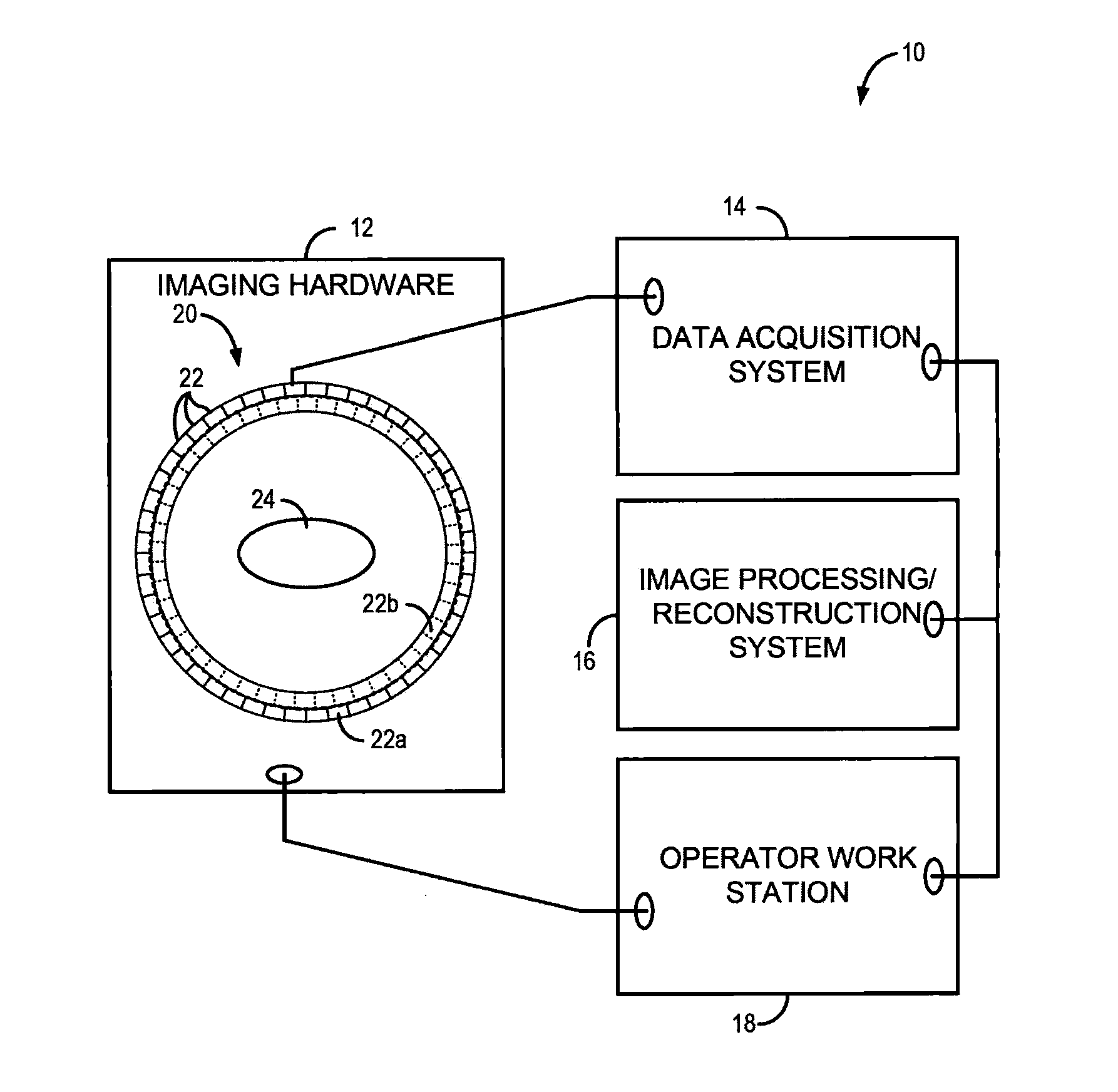
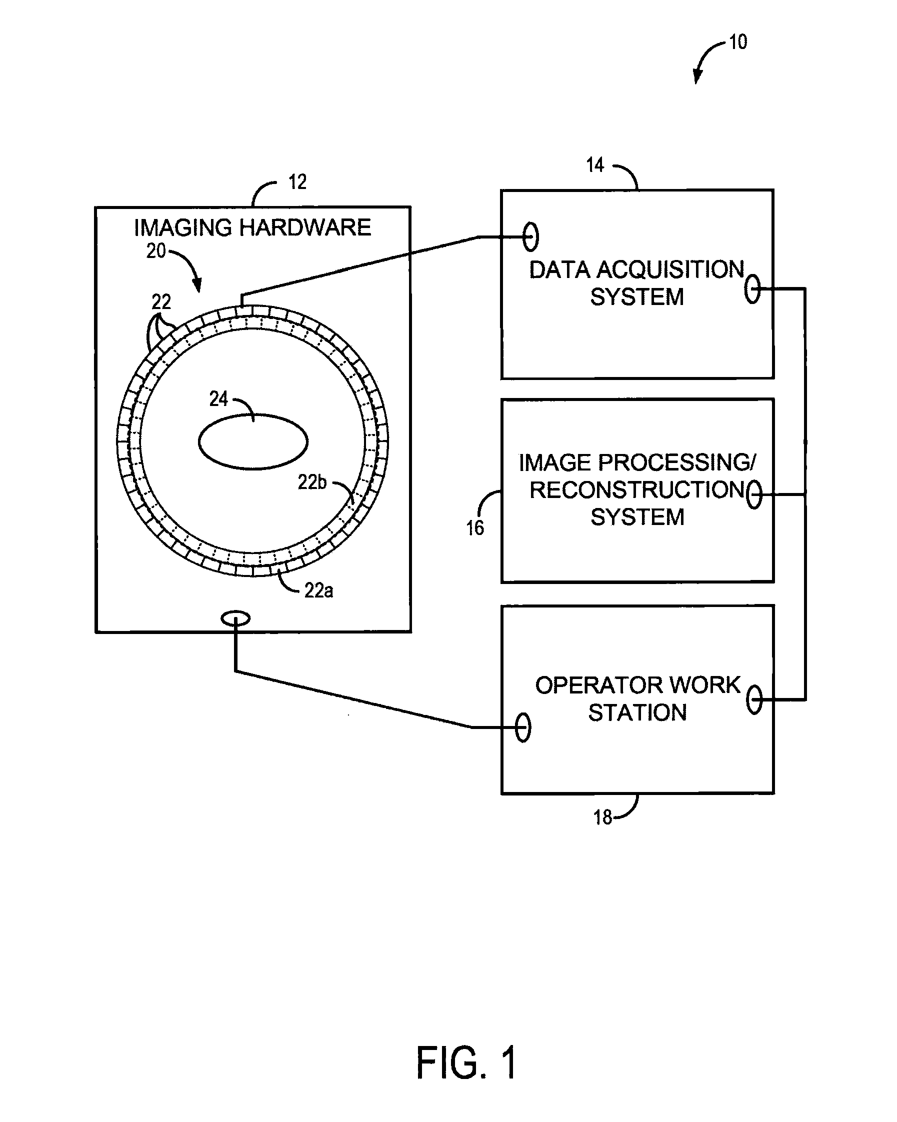
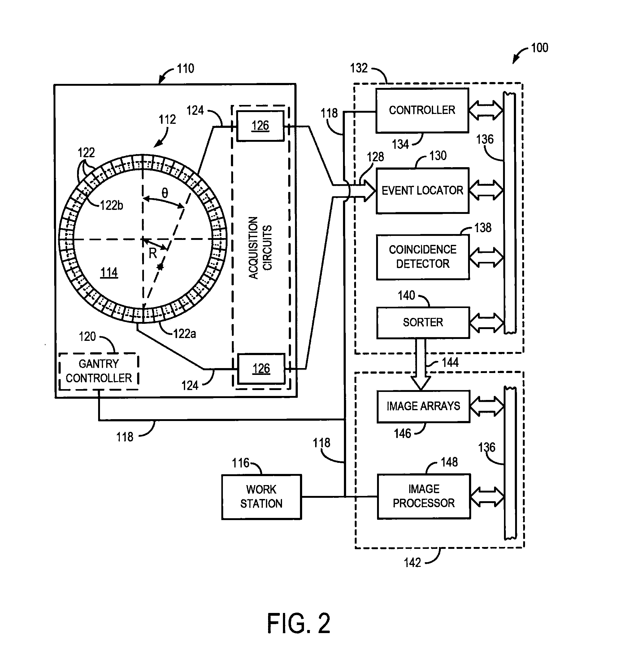
[0026]The present invention recognizes that one of the great strengths of emission tomography, such as positron emission tomography (PET), is the ability to obtain images with high spatial resolution and sensitivity using any of a number of molecular or physiologic targets and different radiotracers. The development of new probes for imaging hypoxia, cell proliferation, blood flow, and numerous other molecular targets, which offers high potential for better diagnosis, characterization of disease, and image-guided personalized medicine. However, much of this potential remains unrealized because current technology permits only one radiotracer to be imaged at a time. Consequently, multiple scanning sessions need to be coordinated, and even scheduled on different days, to obtain complementary information from multiple radiotracers, which results in high costs, image alignment issues, and a long and onerous experience for the patient. As will be described, the present invention overcomes...


PUM


Abstract

A method and system for acquiring a series of medical images during a common imaging process, includes a plurality of detectors configured to be arranged to acquire gamma rays emitted from a subject as a result of multiple radiotracers administered to the subject and communicate signals corresponding to acquired gamma rays. A data processing system is configured to receive the signals from the plurality of detectors and identify temporal information and energy information of photons of the acquired gamma rays. A reconstruction system is configured to receive the signals, the temporal information, and the energy information from the data processing system and reconstruct therefrom a series of medical images of the subject, wherein at least one of the images in the series of medical images corresponds to only to information acquired from gamma rays emitted as a result of a given one of the multiple radiotracers.

Description

CROSS-REFERENCE TO RELATED APPLICATIONS[0001]This application is based on, claims priority to, and incorporates herein by reference in its entirety, U.S. Provisional Application Ser. No. 61 / 640,292, filed Apr. 30, 2012, and entitled “MULTIPLEXABLE EMISSION TOMOGRAPHY”STATEMENT REGARDING FEDERALLY SPONSORED RESEARCH[0002]N / ABACKGROUND OF THE INVENTION[0003]The present invention relates to systems and methods for emission tomography and, more particularly, to systems and methods for multiplexable emission tomography that allow researchers and clinicians to acquire data from multiple, distinguishable sources using emission tomography.[0004]There are a variety of emission tomography imaging systems and methods. For example, single photon emission computed tomography (SPECT) and positron emission tomography (PET) are two clinically important examples. Both PET and SPECT, and other various emission tomography systems and methods, utilize a radionuclide, typically bonded or otherwise coupl...

Claims


Application Information

Patent Timeline
Patent Type & Authority Applications(United States)
IPC IPC(8): G01T1/29A61B6/00A61B6/03
CPCG01T1/2985A61B6/4258A61B6/5205A61B6/037
Inventor LAGE, EDUARDO M.HERRAIZ, JOAQUIN L.PAROT, VICENTE J.
Owner MASSACHUSETTS INST OF TECH
Why Eureka
Try Eureka