Method for non-invasive quantitative assessment of radioactive tracer levels in the blood stream

a radioactive tracer and blood stream technology, applied in the field of non-invasive quantitative assessment of radioactive tracer levels in the blood stream, can solve the problems of difficult interpretation of conventional pharmacokinetic modelling by dynamic scanning or blood withdrawal, and it takes a long time for the underlying pharmacokinetics to reach

Inactive Publication Date: 2012-09-13
NETHERLANDS CANCER INST
View PDF8 Cites 11 Cited by
  • Summary
  • Abstract
  • Description
  • Claims
  • Application Information

AI Technical Summary

Benefits of technology

[0016]Because the method allows for quantitative monitoring of fast changes in tracer concentration, pharmacokinetics can be assessed while the uptake and clearance of tracer in the human system does not yet occur at equilibrium rates. This is a further major advantage over the currently existing methodologies, which require the pharmacokinetic system to be in equilibrium to assess pharmacokinetic rates of tracer transport. Especially when new antibody-labelled tracers are used, it will take a long time for the underlying pharmacokinetics to reach equilibrium state, thus rendering conventional pharmacokinetic modelling by dynamic scanning or blood withdrawal difficult to interpret. In the method provided by the present invention it would be feasible for patients to wear a small portable scanner for days if necessary.

Problems solved by technology

Especially when new antibody-labelled tracers are used, it will take a long time for the underlying pharmacokinetics to reach equilibrium state, thus rendering conventional pharmacokinetic modelling by dynamic scanning or blood withdrawal difficult to interpret.

Method used


Image

  • Method for non-invasive quantitative assessment of radioactive tracer levels in the blood stream
  • Method for non-invasive quantitative assessment of radioactive tracer levels in the blood stream
  • Method for non-invasive quantitative assessment of radioactive tracer levels in the blood stream

Examples


experiment 1

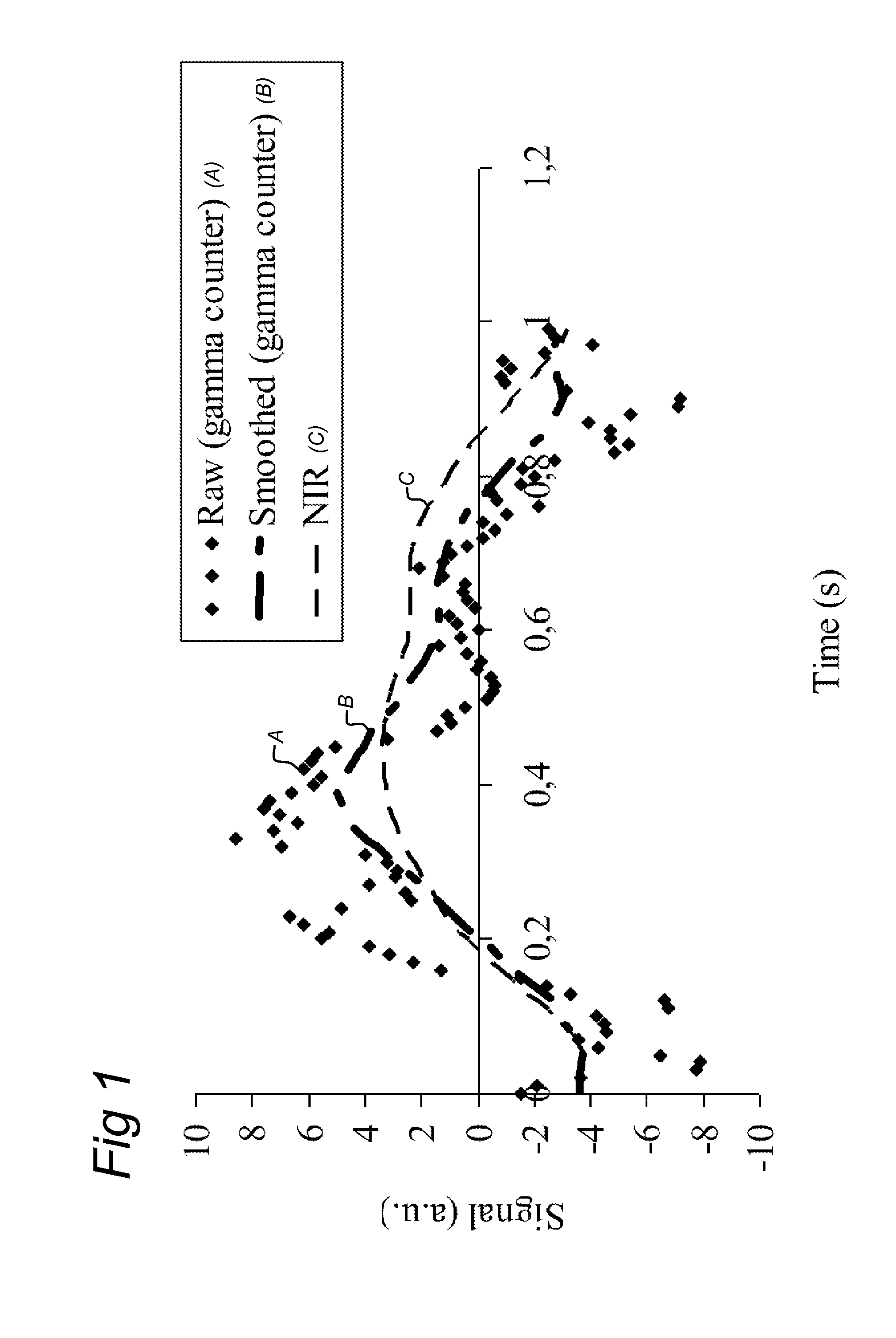
[0055]Arterial vessels vary in diameter due to the pumping movement of the heart. When radiotracers are injected intravenously the radiotracers will dissolve in the blood stream. A gamma counter placed over a part of the body containing arterial vessels will measure a time varying volume of arterial blood. The concentration of radiotracer is assumed to be constant within one heartbeat, therefore causing the number of counts to vary with the changes in the volume of the arterial vessels. In addition, due to the redistribution of the radiotracer in other organs the concentration radiotracer in the blood will decrease. This decrease is reflected in a diminishing of the amplitude of the heartbeat-induced volume changes.

[0056]A change in the amount of circulating radioactive tracer is measured using phase-sensitive detection of the time-varying signal and the changing arterial blood volume. To validate that the time-varying signal is indeed caused by volume changes of the arterial blood ...

experiment 2

[0061]Fluorine-18 labelled fluoroazomycin arabinoside ([18H]FAZA), has been developed as an PET tracer for assessment of Tumour hypoxia. For evaluation of the distribution over time of this tracer dynamic Pet scans have been performed during the first 20 minutes after administration of the tracer. Patients were measured twice in two consecutive days. To evaluate the Phase sensitive detector, we placed the probe between de knees of the patient while the dynamic PET scan was performed. The dynamic scan was constructed in time frames with a duration of 2 seconds. One ROI was placed over the left carotic artery an other ROI over an adjacent muscle.

[0062]FIGS. 4A and 4C show the phase sensitive signal acquired in the knee as compared to the measurement acquired with the PET from the carotic artery. FIGS. 4B and 4D show the low pass filtered signal from the probe compared to the PET signal from the musculature in the neck.


PUM


Abstract

A method and system for non-invasive quantitative assessment of radionuclide tracer levels in the blood stream. The method relies on the finding that the gamma-radiation signal acquired using a gamma scintillation probe, is the resultant from a wave-component with changing amplitude, resulting from the radiotracer in the bloodstream and a non-wave background component resulting from radiotracer distributed throughout the tissue surrounding the arterial vessel. The method involves phase-sensitive conversion of the ‘input signal’, extracting there from an output signal representing the signal component originating from the bloodstream, using the changes in arterial blood volume as the reference wave-form. A particular aspect of the invention concerns methods of quantitative PET or SPECT imaging wherein the concentration of radionuclides in the blood stream as a function of time after injection is assessed using the method of the invention.

Description

FIELD OF THE INVENTION[0001]The present invention concerns a method for non-invasive quantitative assessment of radionuclide tracer levels in the blood stream, as well as the system for use in such methods. A particular aspect of the invention concerns methods of quantitative PET or SPECT imaging wherein the concentration of radionuclides in the blood stream as a function of time after injection is assessed using the method of the invention.BACKGROUND OF THE INVENTION[0002]Positron emission tomography (PET) scanning and single photon emission computed tomography (SPECT) are diagnostic tools for non-invasively imaging living organisms, essential to the investigation of chemical and functional processes in biochemistry, biology, physiology, anatomy, molecular biology, and pharmacology. While techniques, such as x-rays, computed tomography (CT), and magnetic resonance imaging (MRI) provide anatomical images, PET and SPECT scanning techniques provide insight into biochemical changes tha...

Claims


Application Information

Patent Timeline
Patent Type & Authority Applications(United States)
IPC IPC(8): A61B6/00
CPCA61B5/02416A61B6/037A61B6/508A61B6/507A61B6/481
Inventor GILHUIJS, KENNETH GEORGE ANTONIUSSINAASAPPEL, MICHIELMULLER, SARA HILLEGONDA
Owner NETHERLANDS CANCER INST
Why Eureka
Try Eureka