Adjuvant incorporation in immunonanotherapeutics

a technology of adjuvant and immunonuclear therapy, applied in the field of adjuvant incorporation in immunonuclear therapy, can solve the problems of not providing such binding to other types of cells, and achieve the effects of reducing the incidence and reducing the severity of one or more symptoms

Active Publication Date: 2011-11-03
MASSACHUSETTS INST OF TECH +2
View PDF3 Cites 36 Cited by
  • Summary
  • Abstract
  • Description
  • Claims
  • Application Information

AI Technical Summary

Benefits of technology

[0008]The ability of the immunofeature surface to target APCs is a key feature that allows the nanocarriers of the invention to deliver immunostimulatory and immunomodulatory agents to B-cells and / or T-cells when administered to a subject. Such delivery allows the inventive nanocarriers to elicit an immune system response, or to enhance an immune system response. In some embodiments, the synthetic nanocarriers of the invention comprise a surface comprising a plurality of moieties in an amount effective to provide a humoral response to the moieties.
[0130]As discussed below, experiments in FIGS. 29 and 33 demonstrate that synthetic nanocarriers that encapsulate a T cell antigen (OVA, ovalbumin protein) or an antigenic peptide (SIINFEKL, derived from ovalbumin) together with covalently bound adjuvant (CL097 or R848) induce potent proliferation of antigen-specific OT-I CD8+ T cells. This T cell subset (also called cytotoxic T cells) only responds to antigens that are presented in the MHC class-I pathway. Exogenous antigens, such as those provided by conventional vaccines, are preferentially presented by MHC class-II molecules that are not recognized by CD8+ T cells. Only certain subsets of dendritic cells (DC) are capable of shunting some of the exogenous antigen they acquire to the MHC class-I pathway, a process called ‘cross-presentation’. Methods and compositions that result in enhanced cross-presentation of vaccine antigens are desirable because they are predicted to result in improved cytotoxic effector responses that would be useful for the prevention and therapy of cancer and infectious microbes. The data in FIGS. 29 and 33 clearly show that exogenous antigens delivered by inventive synthetic nanocarriers can be cross-presented in vivo resulting in CD8 T cell proliferation in lymph nodes, even when the synthetic nanocarriers do not present an immunofeature surface. However, results shown in FIG. 38 show that synthetic nanocarrier targeting to lymph node DC can be substantially enhanced (at least 5-fold) when synthetic nanocarriers incorporate an immunofeature surface, such as an immunofeature surface comprising nicotine. This gain in targeting would be expected to result in a proportional increase in the amount of antigen and adjuvant that will be available to the DC for cross-presentation to CD8+ T cells. Thus, immunofeature surface-mediated targeting of synthetic nanocarriers would be expected to boost the capacity of synthetic nanocarrier vaccines to elicit cytotoxic T cell responses above and beyond that of synthetic nanocarrier vaccines lacking an immunofeature surface.

Problems solved by technology

In some embodiments, nanocarriers comprising an immunofeature surface exhibit specific low affinity high avidity binding to APCs, and do not provide such binding to other types of cells.

Method used


Image

  • Adjuvant incorporation in immunonanotherapeutics
  • Adjuvant incorporation in immunonanotherapeutics
  • Adjuvant incorporation in immunonanotherapeutics

Examples


example 1

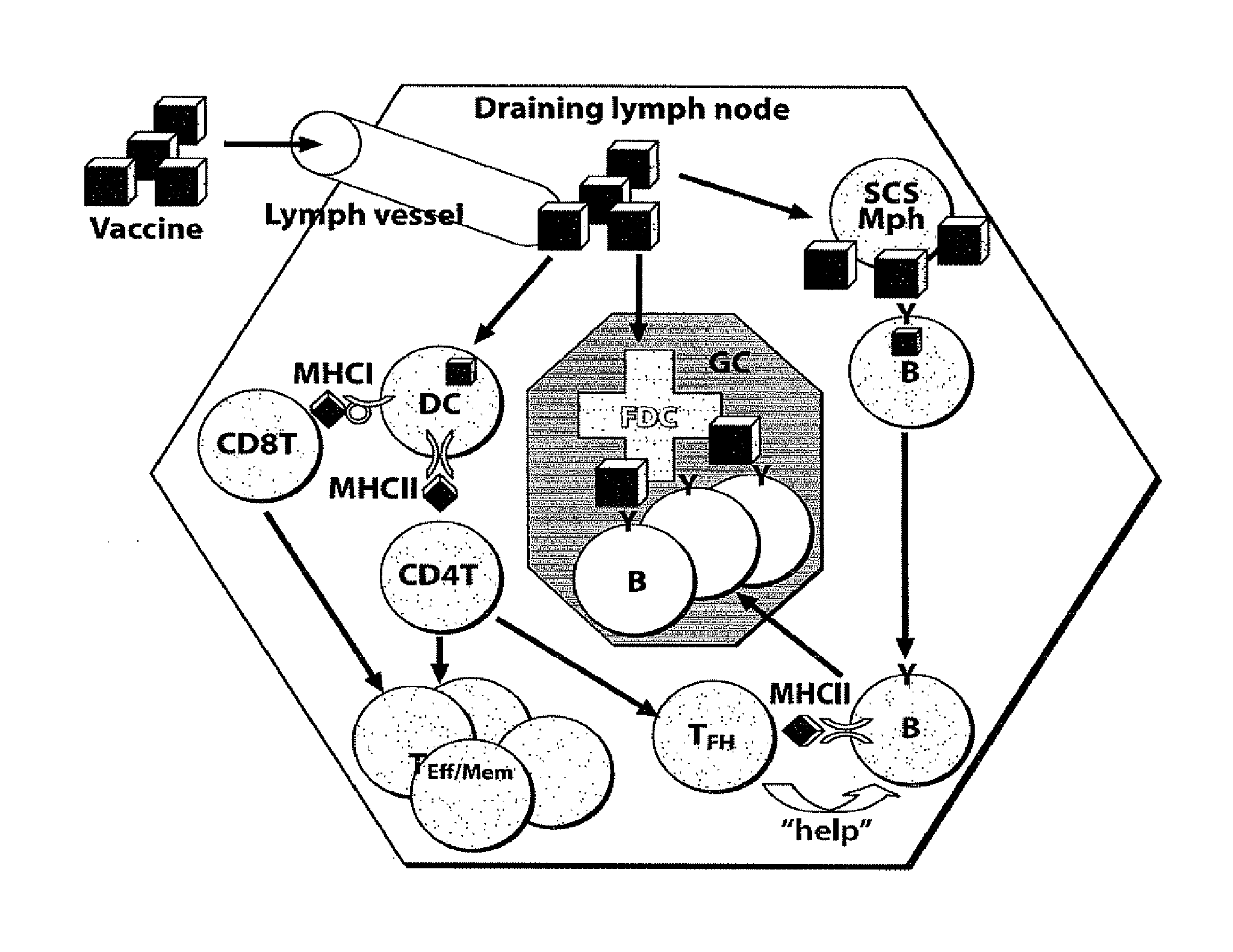
Subcapsular Sinus Macrophages in Lymph Nodes Clear Lymph-Borne Viruses and Present them to Antiviral B Cells

Materials and Methods

[0632]Method Summary

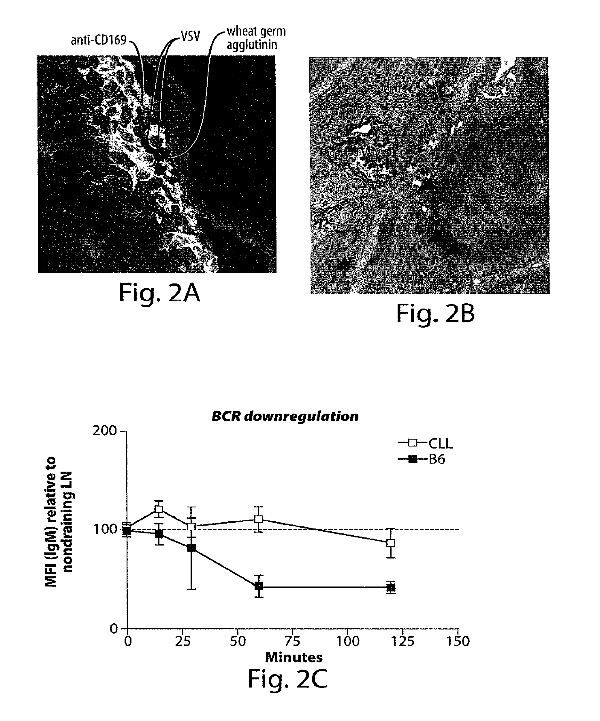
[0633]VSV-IND and VSV-NJ virions were purified from culture supernatants of infected BSRT7 cells and used either unmodified or fluorescently labeled with Alexa-568 (red) or Alexa-488 (green). Fluorescent viruses used for tissue imaging were UV-irradiated to prevent generation of non-fluorescent progeny. Fluorescent labeling or UV-irradiation of VSV-IND particles did not affect their antigenicity or their ability to elicit a calcium flux in VI10YEN cells (not shown). Following fluorescent virus injection into footpads, draining popliteal LNs were harvested for analysis by electron microscopy or to generate frozen sections for immunostaining and confocal microscopy. To image adoptively transferred B cells in LNs, VI10YEN and wildtype B cells were fluorescently labeled and co-transferred by i.v. injection into wildtype or mutant recipient ...

example 2

Exemplary Lipid-Based Vaccine Nanotechnology Architectures

[0671]Liposome Nanocarriers

[0672]In some embodiments, small liposomes (10 nm-1000 nm) are manufactured and employed to deliver, in some embodiments, one or multiple immunomodulatory agents to cells of the immune system (FIG. 3). In general, liposomes are artificially-constructed spherical lipid vesicles, whose controllable diameter from tens to thousands of nm signifies that individual liposomes comprise biocompatible compartments with volume from zeptoliters (10−21 L) to femtoliters (10−15 L) that can be used to encapsulate and store various cargoes such as proteins, enzymes, DNA and drug molecules. Liposomes may comprise a lipid bilayer which has an amphiphilic property: both interior and exterior surfaces of the bilayer are hydrophilic, and the bilayer lumen is hydrophobic. Lipophilic molecules can spontaneously embed themselves into liposome membrane and retain their hydrophilic domains outside, and hydrophilic molecules ...

example 3

In Vivo Targeting of SCS-Mph Using Fc Fragments from Human IgG

[0690]Fluorescent unmodified control nanoparticles (top panel, FIG. 24A) or Fc surface-conjugated targeted nanoparticles (middle and lower panel, FIG. 24A) were injected into footpads of anesthetized mice, and the draining popliteal lymph node was excised 1 hour later and single-cell suspensions were prepared for flow cytometry. Targeted nanoparticles were also injected into mice one week after lymph node macrophages had been depleted by injection of clodronate-laden liposomes (lower panel, FIG. 24A). The cell populations in gates were identified as nanoparticle-associated macrophages based on high expression of CD11b. These results indicate that (i) nanoparticle binding depends on the presence of clodronate-sensitive macrophages and (ii) targeted nanoparticles are bound to twice as many macrophages as control nanoparticles.

[0691]The Panels on the right of FIG. 24 show fluorescent micrographs of frozen lymph node sections...


PUM

PropertyMeasurementUnit
timeaaaaaaaaaa
diameteraaaaaaaaaa
densityaaaaaaaaaa

Abstract

The present invention provides compositions and systems for delivery of nanocarriers to cells of the immune system. The invention provides nanocarriers capable of stimulating an immune response in T cells and / or in B cells. The invention provides nanocarriers that comprise an immunofeature surface and an immunostimulatory moiety. In some embodiments, the immunostimulatory moiety is an adjuvant. The invention provides pharmaceutical compositions comprising inventive nanocarriers. The present invention provides methods of designing, manufacturing, and using inventive nanocarriers and pharmaceutical compositions thereof.

Description

RELATED APPLICATIONS[0001]This application is a continuation-in-part of, and claims priority to, PCT Application Serial No. PCT / US08 / 011,932, filed Oct. 12, 2008. This application also claims priority to U.S. patent application Ser. No. 12 / 428,395, filed Apr. 22, 2009. The entire contents of the aforementioned applications are incorporated herein by reference.GOVERNMENT SUPPORT[0002]This invention was made with government support under Grant Nos. EB003647, AI072252, and AI069259, awarded by the NIH. The government has certain rights in this invention.BACKGROUND OF THE INVENTION[0003]Many current vaccines against microbial pathogens comprise live attenuated or non-virulent strains of the causative microorganisms. Many vaccines comprise killed or otherwise inactivated microorganisms. Other vaccines utilize purified components of pathogen lysates, such as surface carbohydrates or recombinant pathogen-derived proteins. Vaccines that utilize live attenuated or inactivated pathogens typic...

Claims


Application Information

Patent Timeline
Patent Type & Authority Applications(United States)
IPC IPC(8): A61K9/14A61K39/39A61K38/21A61P37/04A61K38/20A61K39/02A61K39/12A61K39/00A61K39/002
CPCA61K39/12A61K45/06A61K39/002A61K38/21A61K9/14A61K39/0002A61K39/39A61K39/05A61K39/08A61K38/20A61K39/00A61P19/02A61P31/00A61P35/00A61P37/00A61P37/02A61P37/04A61P37/06A61P43/00
Inventor ALEXIS, FRANKIANNACONE, MATTEOSHI, JINJINBASTO, PAMELAMOSEMAN, ELLIOTT ASHLEYVON ANDRIAN, ULRICHLANGER, ROBERT S.FAROKHZAD, OMID C.TONTI, ELENA
Owner MASSACHUSETTS INST OF TECH
Why Eureka
Try Eureka