Registering mr patient data on the basis of generic models

a generic model and patient data technology, applied in image data processing, diagnostics, sensors, etc., can solve the problems of x-ray radiation used to generate images being a burden on patients' health, bone structures, and generally not being identified or identifiable in mr recordings, so as to enhance navigation accuracy and accurate detection and adjustment of model positions

Active Publication Date: 2011-11-03
BRAINLAB
View PDF0 Cites 32 Cited by
  • Summary
  • Abstract
  • Description
  • Claims
  • Application Information

AI Technical Summary

Benefits of technology

[0017] An advantage of the method is that when a generic or statistical model is used that has been adapted to the patient, it is no longer necessary, for a treatment in which medical navigation is to be provided, to produce a separate data set for the body structure. On the one hand, this spares the patient a high radiation load, for example from recording numerous x-ray or CT images, and on the other hand, the cost of producing such data sets can be minimized. Further, linking the generic body structure data with patient-characteristic detection data provides a data set that can be used to provide very accurate medical navigation. The generic model, which can be a universal model for the relevant body structure that includes all relevant data, does not from the outset comprise any data that are specifically tailored to the relevant patient. The generic model, however, once adapted with the aid of patient-characteristic detection data, does comprise sufficient anatomical or body structure data to provide a sufficiently accurate basis for medical navigation.
[0028] The generic model thus can be generated exclusively from or by means of CT reference data sets, such that the generic model can be accurately and quickly adapted to the patient-specific fluoroscopic images. The generic model can be generated from the plurality of CT reference data sets, such as CT training reference data sets, and the CT main shape reference data set. One MR main shape reference data set preferably is registered to the CT main shape reference data set, wherein a correlation exists between the generic model and the MR main shape reference data set.
[0034] This approach exhibits an array of advantages. Non-patient-specific CT data sets that are easy to ascertain or record can be used to produce the generic model. In this approach, only the main shape reference data set comprises an MR main shape reference data set and a CT main shape reference data set, which results in a significant reduction in labor and costs. Also, only the data of the main shape reference data set that also can be displayed in the fluoroscopic images and / or the generic model, and the patient-specific position of which can thus be correctly ascertained to a high probability, may be changed based on the ascertained transformation protocol. The fixed registration of the adapted main shape reference data set to the patient-specific MR data set also represents a simple, accurate and quick process.
[0037] It is, however, also possible to produce individual x-ray images of the patient even during the treatment and to then incorporate this information into adapting the generic model. An advantage as compared to conventional “x-ray navigation” is that it is not necessary to produce a large number of x-ray images, as used in x-ray image based navigation. By contrast, it is sufficient to produce only one or very few x-ray images in order to adapt the generic model, which in addition can be limited to a very small portion of the body. This significantly reduces the radiation load as compared to conventional x-ray navigation.
[0045] The generic model thus can be fused with patient-specific information or image data either automatically, for example by automatically identifying particular anatomical features that are critical for fusing, or also manually, for example by shifting, rotating, and / or stretching / warping. If the generic model is fused with actual patient information with the aid of acquiring an indefinite number of items of point information on the patient (landmarks), it is possible to use a so-called surface matching method, e.g., a computer-assisted image adapting method, to fuse the image data. Detecting the diagnostic data and adapting the generic model from the various methods described above can be combined such that in addition to the diagnostic data (for example, intra-operatively acquired x-ray images), additional points on the patient also are recorded in the form of landmarks or randomly acquired points and used to accurately detect and adjust the position of the model or even its shape, so as to enhance navigation accuracy.

Problems solved by technology

A drawback to such methods, however, is that x-ray radiation used to generate the images can be a burden to the patient's health.
Bone structures, however, generally are poorly identified or not identifiable in MR recordings.
This, however, can incur significant costs since computer tomographs and magnetic resonance tomographs are very expensive both to purchase, maintain and operate.
Further, a plurality of CT recordings typically are produced, which can place a high radiation load on the patient.
However, such systems can lack the required accuracy for the patient to be treated in each case.
These generic models, however, are not primarily based on MR data sets.
Therefore, generic MR data cannot be correspondingly deformed and registered to the fluoroscopic images.

Method used


Image

  • Registering mr patient data on the basis of generic models
  • Registering mr patient data on the basis of generic models
  • Registering mr patient data on the basis of generic models

Examples


Embodiment Construction

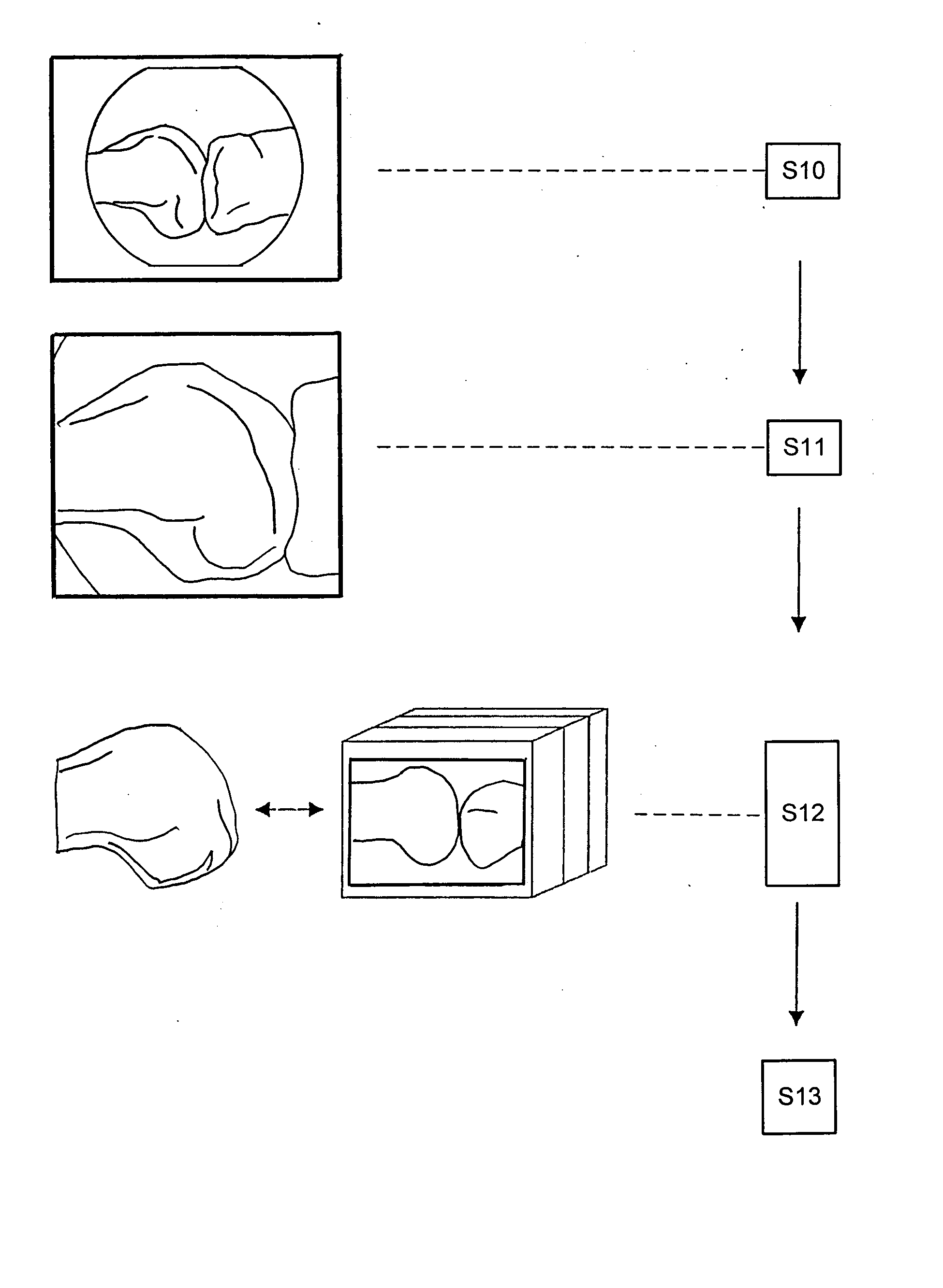
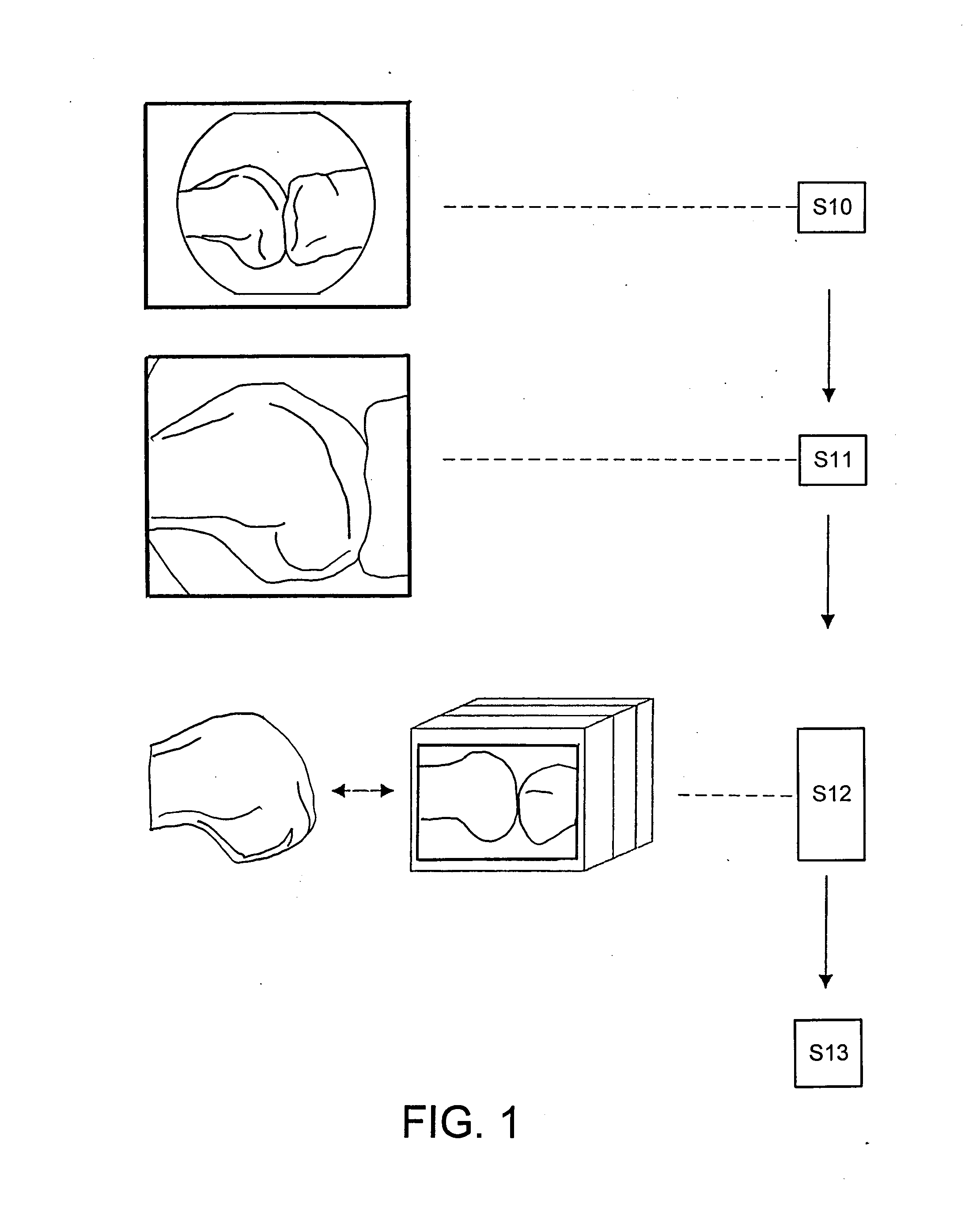
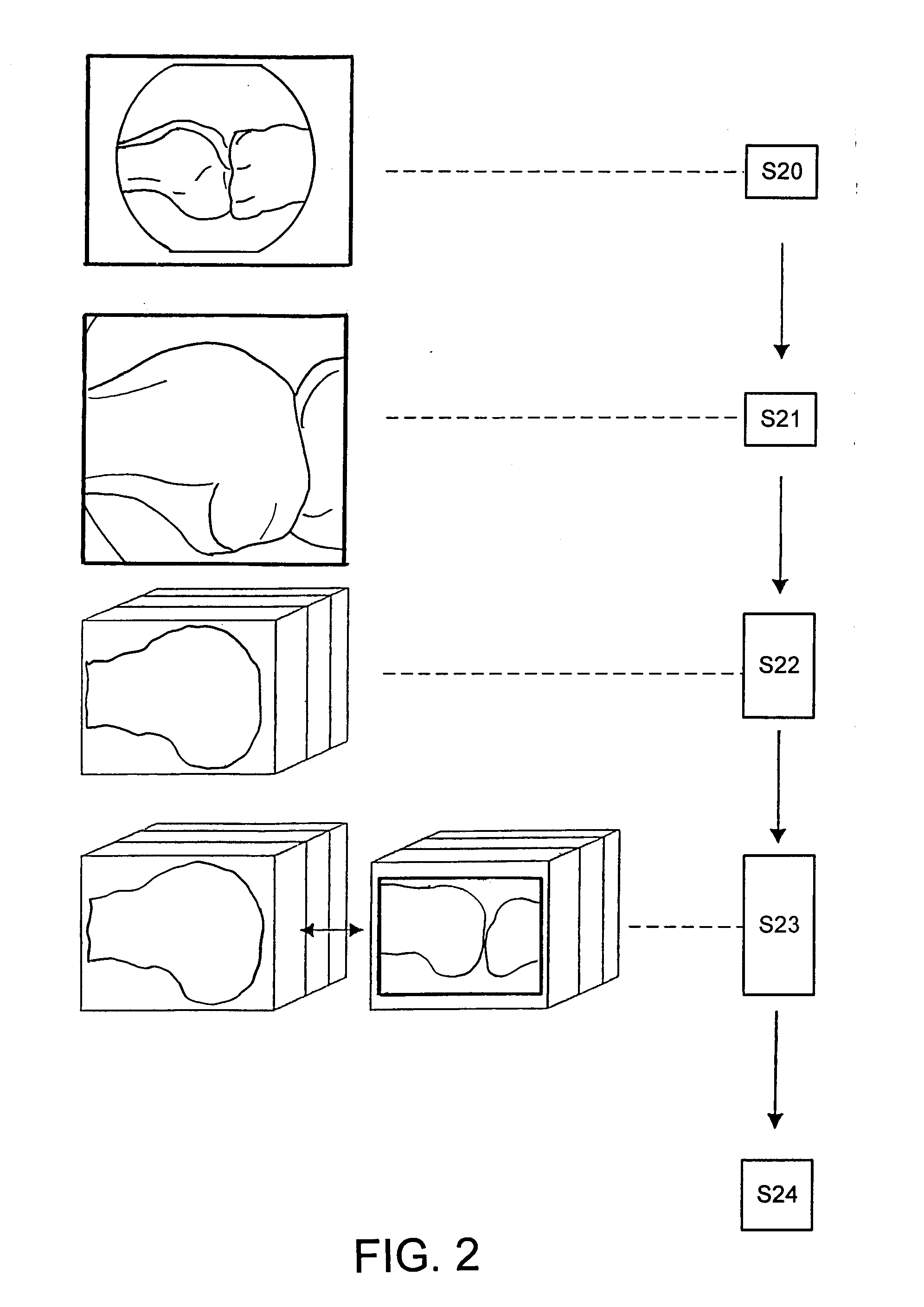
[0055]FIG. 1 is a flow diagram illustrating a first exemplary method for registering patient data on the basis of generic models. At least two fluoroscopic images of a body region of a patient are initially recorded in step S10. These fluoroscopic images contain patient-specific data such as patient-specific structures or shapes. A non-patient-specific adaptive generic model, which, for example, can include a plurality of data sets such as CT data sets, MR data sets, x-ray data sets or other data sets, is adapted to the fluoroscopic image data in step S11 by means of a transformation protocol. The transformation protocol at least partially adapts the initially non-patient-specific generic model to the actual patient-specific structures apparent from the fluoroscopic images. In the next step, step S12, the generic model, which has already been partially adapted, is registered with respect to a patient-specific MR data set, or conversely, the patient-specific MR data set is registered...


PUM


Abstract

A method for registering a non-patient-characteristic three-dimensional magnetic resonance data set (MR data set) to patient-characteristic data includes: producing or providing a non-patient-characteristic three-dimensional generic model of a body or body part containing body structure data; ascertaining or providing two-dimensional patient-characteristic detection data of a patient; using a transformation protocol for data-linking the body structure data of the three-dimensional generic model to the two-dimensional patient-characteristic detection data to change or adapt the generic model of the body or body part based on the ascertained two-dimensional patient-characteristic detection data, wherein the three-dimensional generic model is at least correlated with a three-dimensional MR reference data set; and changing or deforming at least a part of the three-dimensional MR reference data set by using the transformation protocol to generate a patient-characteristic three-dimensional MR data set that is registered to the fluoroscopic images.

Description

RELATED APPLICATION DATA [0001] This application claims priority of U.S. Provisional Application No. 60 / 822,706 filed on Aug. 17, 2006, which is incorporated herein by reference in its entirety.FIELD OF THE INVENTION [0002] The present invention relates to a method and device for registering a non-patient-characteristic three-dimensional magnetic resonance (MR) data set to patient-characteristic image data, and more particularly, to at least two fluoroscopic images of the patient. Based on these image data, it is then possible to perform computer-assisted medical navigation. BACKGROUND OF THE INVENTION [0003] When examining a patient or preparing for surgery, in particular surgery in the region of bones such as, for example, spine, hip joint or knee operations, x-ray recordings or computer tomography (CT) recordings of the affected body structure are taken. From these recordings, the body structures can be clearly displayed. A drawback to such methods, however, is that x-ray radiati...

Claims


Application Information

Patent Timeline
Patent Type & Authority Applications(United States)
IPC IPC(8): A61B5/055
CPCA61B19/50A61B19/52A61B19/5244A61B2017/00712A61B2019/505G06T2207/30004A61B2019/5238A61B2019/5255A61B2019/5289A61B2019/566G06T7/0024A61B2019/5236A61B90/36A61B2034/2055A61B2034/256A61B34/20A61B2090/364A61B2090/376A61B34/10A61B2034/105A61B2090/374G06T7/30
Inventor FEILKAS, THOMASSCHAFFRATH, CLAUS
Owner BRAINLAB
Why Eureka
Try Eureka