System and method for image-based respiratory motion compensation for fluoroscopic coronary roadmapping

a fluoroscopic coronary roadmapping and motion compensation technology, applied in image enhancement, image analysis, instruments, etc., can solve the problems of respiratory motion being less reproducible, difficult to compensate breathing motion, motion compensation, etc., and achieve large image motion and reliable estimation of soft tissue motion.

Inactive Publication Date: 2011-11-10
SIEMENS HEALTHCARE GMBH
View PDF8 Cites 24 Cited by
  • Summary
  • Abstract
  • Description
  • Claims
  • Application Information

AI Technical Summary

Benefits of technology

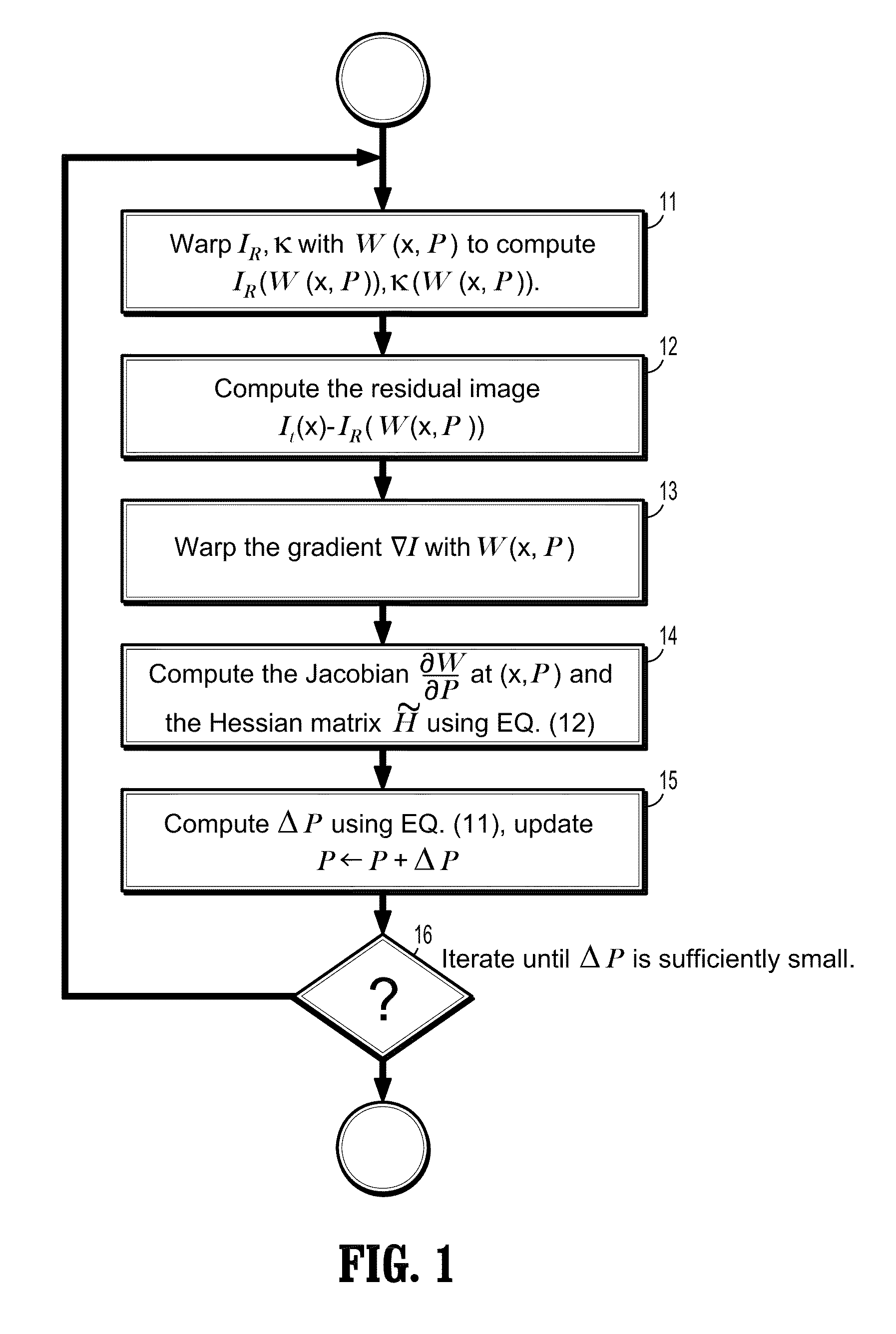
[0006]Exemplary embodiments of the invention as described herein generally include methods and systems for image-based respiratory motion compensation for coronary roadmapping in fluoroscopic images. Static structures can be identified in fluoroscopic images over a cardiac cycle, which allows image information of soft tissues to be separated and used for respiratory motion estimation. An extended Lucas-Kanade algorithm involving a weighted sum-of-squared-difference (WSSD) measure can estimate soft tissue motion reliably when a layer of static structures is present. A temporally compositional motion model can handle large image motion incurred by deep breathing. The recovered image motion allows dynamically moving the coronary roadmap to match the live fluoroscopic images when contrast medium has disappeared.

Problems solved by technology

Motion compensation is an important issue for image guided coronary angioplasty procedures.
In contrast, respiratory motion is less reproducible and drawbacks are associated with respiratory gating.
It remains a challenging issue to compensate breathing motion in live fluoroscopy, which consists of transparent layers of static bone tissues and moving soft tissues as well as contrast-filled vessels and devices.
In cardiac fluoroscopic images, transparent layers of both static bone tissues and soft tissue structures undergoing cardiac and respirator motion are present, making image motion compensation a challenging task.
The performance of such an approach is limited when processing images with poor guidewire visibility or with multiple wires in the vicinity.

Method used


Image

  • System and method for image-based respiratory motion compensation for fluoroscopic coronary roadmapping
  • System and method for image-based respiratory motion compensation for fluoroscopic coronary roadmapping
  • System and method for image-based respiratory motion compensation for fluoroscopic coronary roadmapping

Examples


Embodiment Construction

[0035]Exemplary embodiments of the invention as described herein generally include systems and methods for motion compensation of respiratory motion in live X-ray fluoroscopic images. Accordingly, while the invention is susceptible to various modifications and alternative forms, specific embodiments thereof are shown by way of example in the drawings and will herein be described in detail. It should be understood, however, that there is no intent to limit the invention to the particular forms disclosed, but on the contrary, the invention is to cover all modifications, equivalents, and alternatives falling within the spirit and scope of the invention.

[0036]As used herein, the term “image” refers to multi-dimensional data composed of discrete image elements (e.g., pixels for 2-D images and voxels for 3-D images). The image may be, for example, a medical image of a subject collected by computer tomography, magnetic resonance imaging, ultrasound, or any other medical imaging system know...


PUM


Abstract

A method for compensating respiratory motion in coronary fluoroscopic images includes finding a set of transformation parameters of a parametric motion model that maximize an objective function that is a weighted normalized cross correlation function of a reference image acquired at a first time that is warped by the parametric motion model and a first incoming image acquired at a second time subsequent to the first time. The weights are calculated as a ratio of a covariance of the gradients of the reference image and the gradients of the first incoming image with respect to a root of a product of a variance of the gradients of the reference image and the variance of the gradients of the first incoming image. The parametric motion model transforms the reference image to match the first incoming image.

Description

CROSS REFERENCE TO RELATED UNITED STATES APPLICATIONS[0001]This application claims priority from “Image-based Soft Tissue Motion Analysis and Motion Compensation in Fluoroscopic Images”, U.S. Provisional Application No. 61 / 313,862 of Zhu, et al., filed Mar. 15, 2010, and “Self-Assessing Image-Based Respiratory Motion Compensation For Fluoroscopic Coronary Roadmapping”, U.S. Provisional Application No. 61 / 408,156 of Manhart, et al., filed Oct. 29, 2010, the contents of both of which are herein incorporated by reference in their entireties.TECHNICAL FIELD[0002]This disclosure is directed to methods for motion compensation of respiratory motion in live X-ray fluoroscopic images.DISCUSSION OF THE RELATED ART[0003]Motion compensation is an important issue for image guided coronary angioplasty procedures. As the primary imaging modality for cardiac intervention, live X-ray fluoroscopy is widely used in percutaneous coronary intervention (PCI) procedures, for instance, for directing guidew...

Claims


Application Information

Patent Timeline
Patent Type & Authority Applications(United States)
IPC IPC(8): G06K9/00
CPCG06T7/204G06T2207/30048G06T2207/10121G06T7/248
Inventor ZHU, YINGTSIN, YANGHAISUNDAR, HARI
Owner SIEMENS HEALTHCARE GMBH
Why Eureka
Try Eureka