Computer Aided Detection Of Abnormalities In Volumetric Breast Ultrasound Scans And User Interface

a breast ultrasound and computer assisted technology, applied in the field of medical imaging systems and processes, can solve the problems of insensitivity of mammography, inability to use gadolinium in contrast agent enhanced mri, and less attractiveness

Inactive Publication Date: 2012-01-19
QVIEW MEDICAL
View PDF19 Cites 84 Cited by
  • Summary
  • Abstract
  • Description
  • Claims
  • Application Information

AI Technical Summary

Benefits of technology

[0009]Accordingly, a computer aided detection method that helps radiologists in searching and interpretation of abnormalities would be very useful. According to some embodiments, a novel CAD system is provided for detection of breast cancer in volumetric ultrasound scans.

Problems solved by technology

However, in dense breasts mammography is known to be insensitive.
The use of gadolinium in contrast agent enhanced MRI may not be acceptable in a screening population.
However, the fact that this screening exam, requiring radiologists to perform by hand and read the images as being generated, is time consuming for radiologists makes it less attractive.
Although the image acquisition with ABUS can generally be performed by technicians, radiologists are still required to read the hundreds of images, thus breast imaging with ABUS can still be relatively time consuming.

Method used


Image

  • Computer Aided Detection Of Abnormalities In Volumetric Breast Ultrasound Scans And User Interface
  • Computer Aided Detection Of Abnormalities In Volumetric Breast Ultrasound Scans And User Interface
  • Computer Aided Detection Of Abnormalities In Volumetric Breast Ultrasound Scans And User Interface

Examples


Embodiment Construction

[0032]The following description provides exemplary embodiments only, and is not intended to limit the scope, applicability, or configuration of the disclosure. Rather, the following description of the exemplary embodiments will provide those skilled in the art with an enabling description for implementing one or more exemplary embodiments. It being understood that various changes may be made in the function and arrangement of elements without departing from the spirit and scope of the invention as set forth in the appended claims.

[0033]Specific details are given in the following description to provide a thorough understanding of the embodiments. However, it will be understood by one of ordinary skill in the art that the embodiments may be practiced without these specific details. For example, systems, processes, and other elements in the invention may be shown as components in block diagram form in order not to obscure the embodiments in unnecessary detail. In other instances, well-...


PUM


Abstract

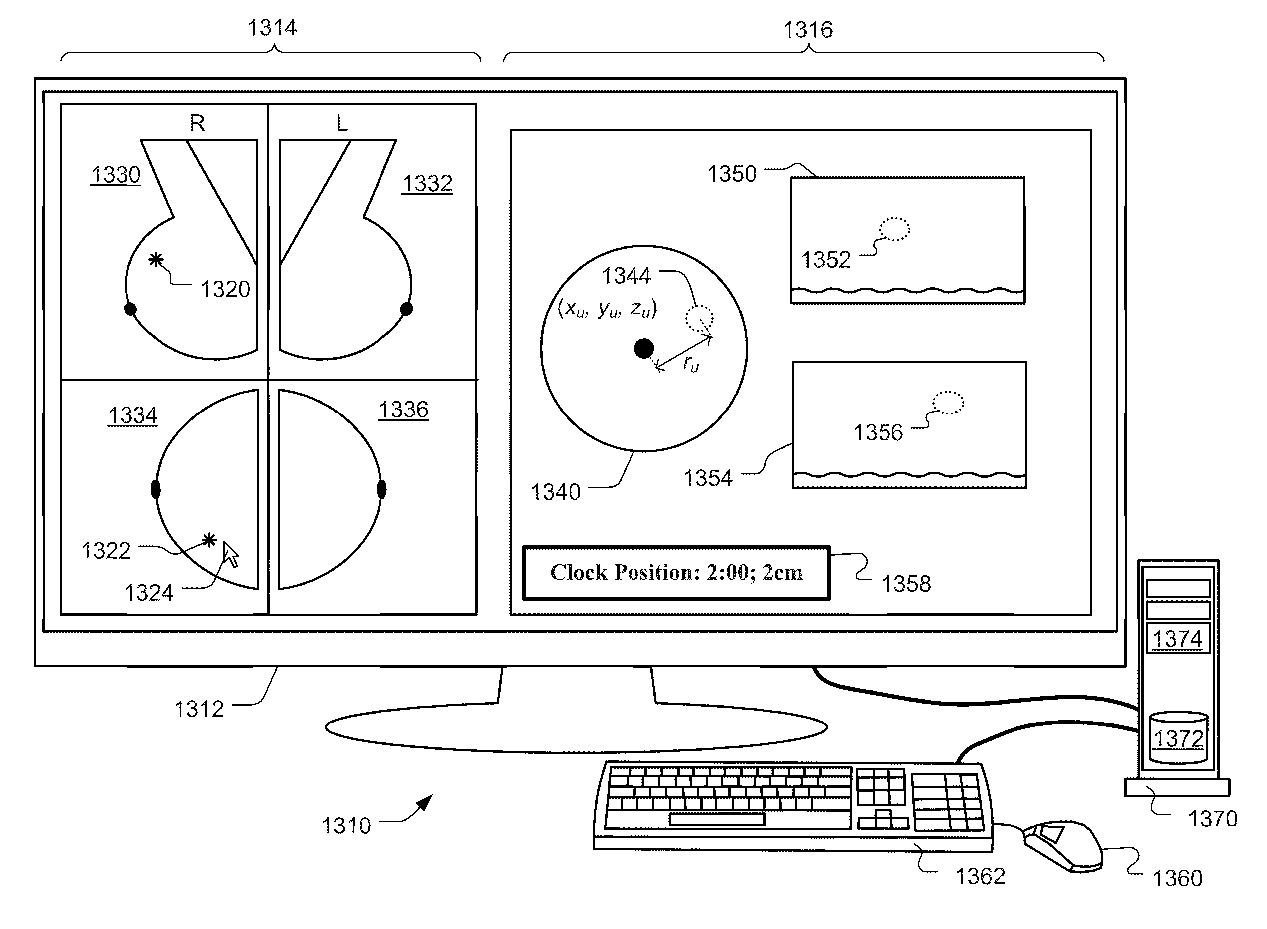
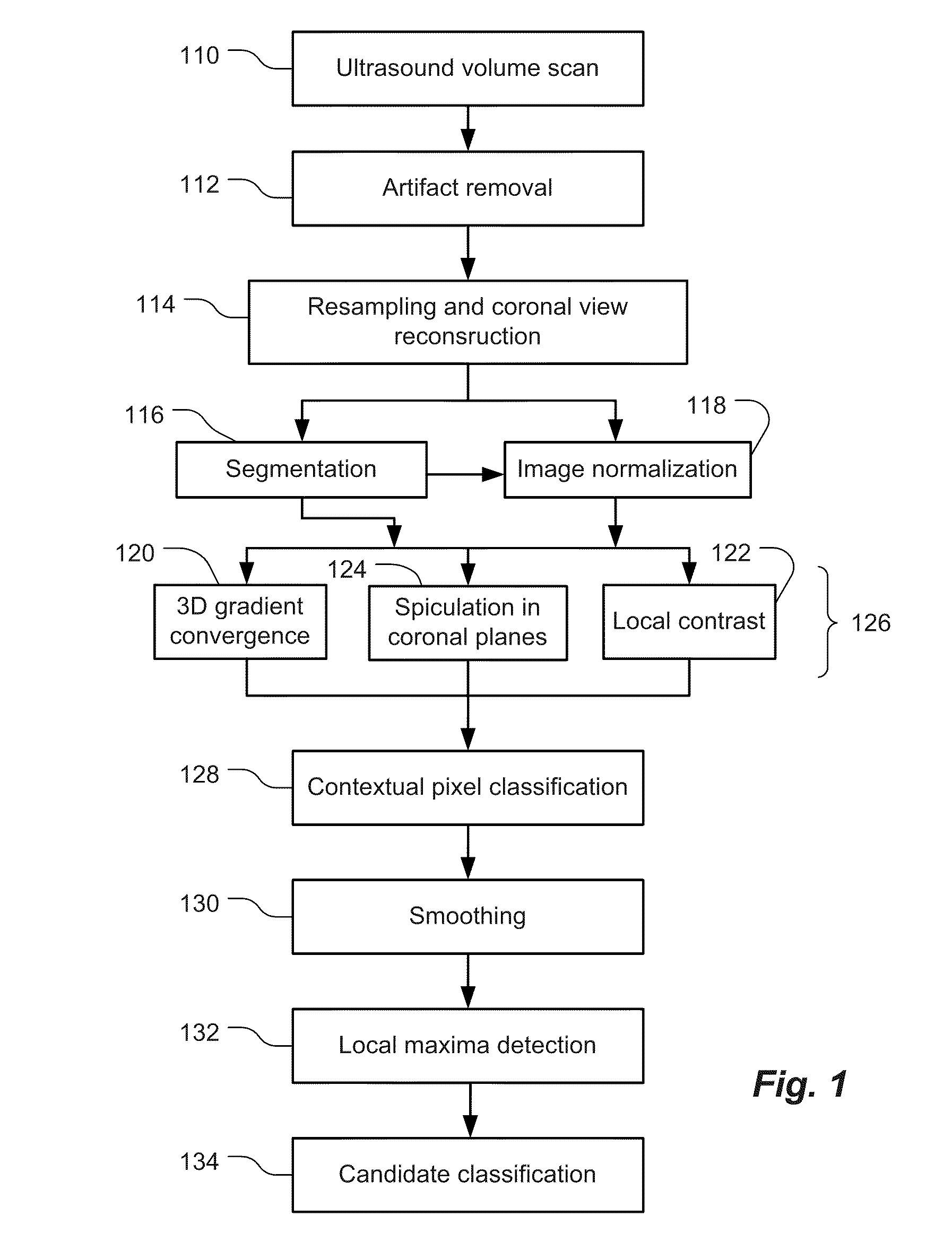
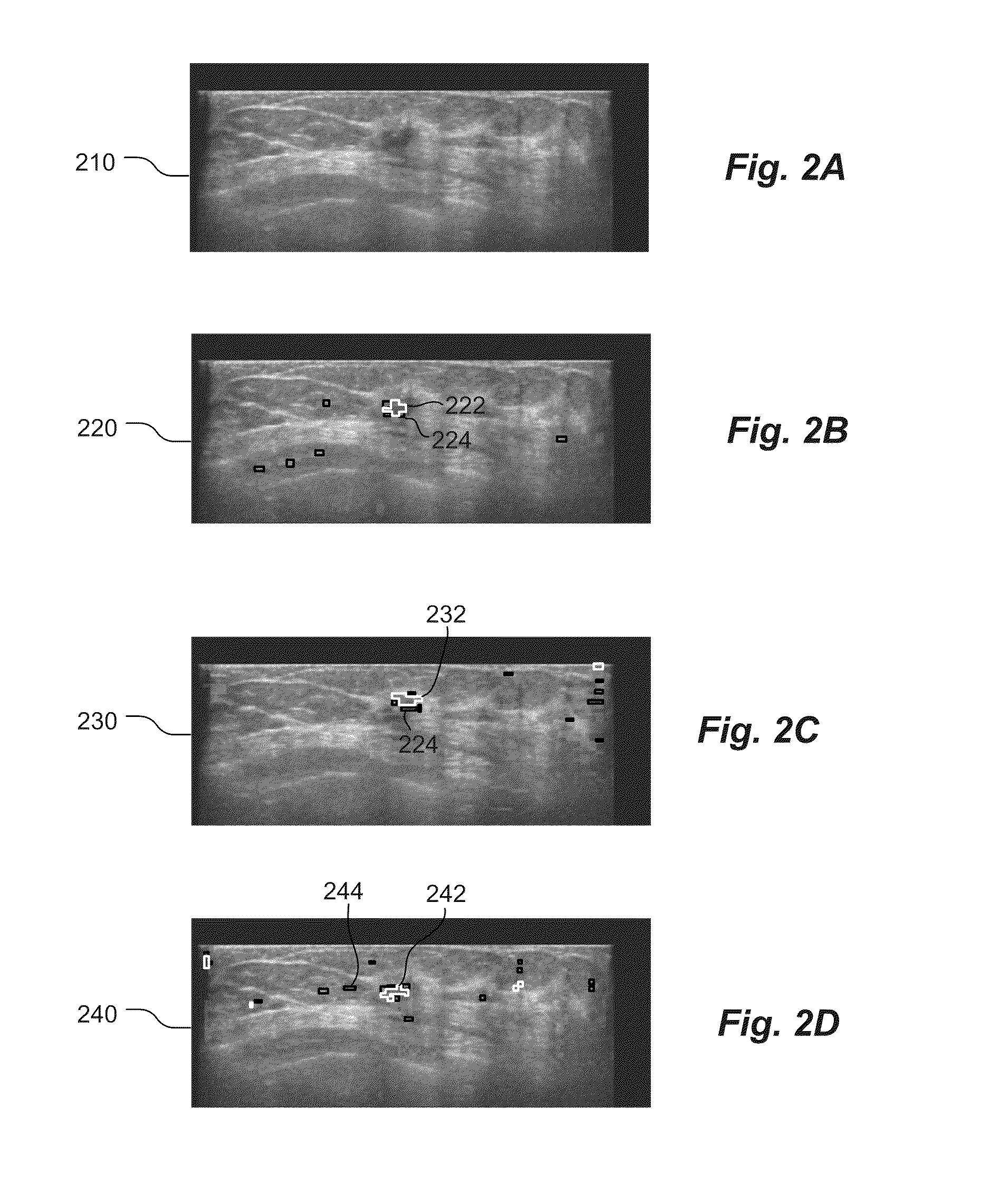
Methods and related systems are described for detection of breast cancer in 3D ultrasound imaging data. Volumetric ultrasound images are obtained by an automated breast ultrasound scanning (ABUS) device. In ABUS images breast cancers appear as dark lesions. When viewed in transversal and sagittal planes, lesions and normal tissue appear similar as in traditional 2D ultrasound. However, architectural distortion and spiculation are frequently seen in the coronal views, and these are strong indicators of the presence of cancer. The described computerized detection (CAD) system combines a dark lesion detector operating in 3D with a detector for spiculation and architectural distortion operating on 2D coronal slices. In this way a sensitive detection method is obtained. Techniques are also described for correlating regions of interest in ultrasound images from different scans such in different scans of the same breast, scans of a patient's right versus left breast, and scans taken at different times. Techniques are also described for correlating regions of interest in ultrasound images and mammography images. Interactive user interfaces are also described for displaying CAD results and for displaying corresponding locations on different images.

Description

BACKGROUND[0001]1. Field[0002]This patent specification relates to medical imaging systems and processes. In particular, the present invention relates to the computer aided detection of breast abnormalities in volumetric breast ultrasound scans, and devices and methods of interactive display of such computer aided detection results.[0003]2. Related Art[0004]Breast cancer screening programs currently use x-ray mammography to find cancers in an early stage when treatment is most effective. However, in dense breasts mammography is known to be insensitive. Therefore, new screening modalities are being investigated that may complement or replace mammography in women with high breast density. The most promising new technologies for screening dense breasts are dynamic contrast enhanced MRI, and automated breast ultrasound scanning. The latter technique is less sensitive than MRI, but has the advantage that it is relatively inexpensive that it does not require the use of a contrast agent. T...

Claims


Application Information

Patent Timeline
Patent Type & Authority Applications(United States)
IPC IPC(8): G06K9/00
CPCG06T7/0012G06T2207/30068G06T2207/10132
Inventor KARSSEMEIJER, NICOZHANG, WEI
Owner QVIEW MEDICAL
Why Eureka
Try Eureka