Detection of fetal cells from maternal blood

a technology of fetal cells and blood, which is applied in the field of detection of fetal cells from maternal blood, can solve the problem of not preventing access

Inactive Publication Date: 2012-03-22
AARHUS UNIV
View PDF1 Cites 6 Cited by
  • Summary
  • Abstract
  • Description
  • Claims
  • Application Information

AI Technical Summary

Benefits of technology

[0025]It is an object of the present invention to provide for a method for detecting foetal cells in a maternal blood sample. The method developed by the inventors is cell type unspecific and enables detection of chromosomes, conservation of cell morphology and immunoassaying of cell type specific proteins.
[0026]Previous methods used for the identification of foetal cells in maternal blood samples were generally aimed at identifying foetal cells of a particular type by including an immunologic selection step selecting specific cell types expected to be present in the maternal blood sample. Foetal cells as trophoblasts, erythroblast (nucleated erythocytes) and foetal leukocytes have been identified using such procedures. The method according to the present invention does not discriminate between different nucleated cell types and thus enables detection of foetal cells of various cell types. The identified cells may thus be any type of foetal nucleated blood cells, such as, but not limited to, leucocytes, stem cells (such as hematopoietic stem cells or mesenchymal stem cells) or placenta derived cells, such as trophoblast cells. The method according to the invention has further provided a method allowing detection of foetal cells of new cell types as the method is not limited to foetal cells of one or more particular cell types. Foetal cells of any cell type may be identified using the method described herein including foetal cells of cell types previously not seen, which may be such as a primitive stem cell.
[0038]Following a re-fixation step may be used to provide further stability and sensibility of the sample during the hybridisation procedures. Such re-fixation step may comprise PFA treatment, such as incubation of the cells in a solution comprising 0.1-5% PFA. Following the samples are conveniently dehydrated in ethanol.
[0052]The method described further enables detection of a rare foetal cell, such as a cell present in a ratio of less than 1 to 103 maternal nucleated cells. Thus the method provided herein enables examination of a large number of cells, which may be performed using an automated scanner system.
[0066]The method according to the invention may further comprise a step of minimizing isolation of binding members recognising non-antigenic material, as may be obtained by protection of the antigenic material and any genetic information encoding the binding member, bound thereto, from agents or treatment capable of rendering the genetic material non-replicable, such as UV irradiation.
[0069]In addition the method of generating binding members according to the invention is not dependent on the specific identity of the antigen, e.g. isolated binding members may recognize different antigens available on / in the target antigenic materiel, e.g. the cell or tissue. The method further allows the generated binding members to be retested on the original target antigenic material whereby specificity may be confirmed.

Problems solved by technology

The method includes applying a mild fixation and permeabilisation step, which at the same time does not prevent accessibility for a hybridisation probes.

Method used


Image

  • Detection of fetal cells from maternal blood
  • Detection of fetal cells from maternal blood
  • Detection of fetal cells from maternal blood

Examples


example 1

Preparation of Peripheral Blood from Pregnant Women

[0254]The maternal blood sample (usually 3 ml) is divided into aliquots of 1 ml. Each aliquot is diluted 1:14 with 0.15 M NaCl, and the cells are allowed to sediment over night at 4° C. After sedimentation, the upper 12 ml of the supernatants are carefully removed and pooled.

[0255]The sedimented cells (total volume 2 ml / tube) are divided into aliquots of 0.5 ml.

Pre-Fixation without Erythrocyte Lysis

[0256]Cells of the supernatant is pre-fixed in a paraformaldehyde solution in PBS, with a final concentration of 0.5% of PFA for 10 minutes.

Recovering and Mounting on Slides

[0257]The cells are recovered by centrifugation for 10 minutes at 500 g. After centrifugation, the supernatant is discarded and the cell pellet is re-suspended in 0.15 M NaCl (usually 20 μl) and smeared onto poly-L-lysine coated slides (usually 2 slides).

Pre-Fixatio with NH4Cl Mediated Erythrocyte Lysis

[0258]Each aliquot of the sedimented cells is diluted with 10 ml of...

example 2

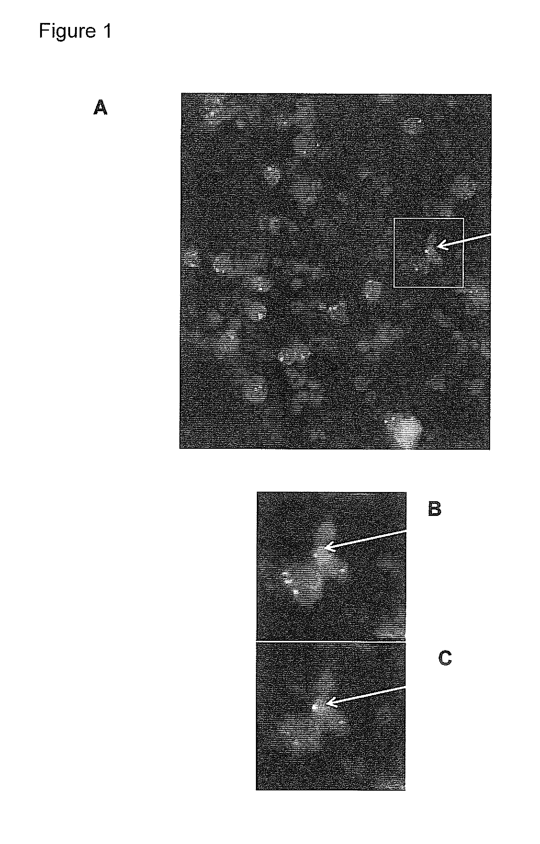
Identification of Male Fetal Cells by Reverse Color Fish and Automated Scanning

[0262]Slides are recovered from the freezer and the airtight plastic back is removed. Before hybridization, slides are fixed and permeabilised 10 minutes in −20° C. cold methanol, 10 minute in −20° C. cold acetone and rinsed in phosphate buffered saline (PBS). Slides are then fixed again for 10 minutes in 2% PBS buffered paraformaldehyde (PFA) and rinsed for 5 minutes in PBS before they are dehydrated 3 minutes each in 60%, 80% and 99.9% ethanol and air-dried.

[0263]Chromosome-specific repeat probes, DXZ1 labeled with spectrum green and DYZ1 Labeled with spectrum orange (Vysis) are used for the first hybridization. Hybridization mixture containing both probes is prepared by mixing 1 part of the X-probe, 1 part of the Y-probe, 1 part of distilled water, and 7 parts of hybridization buffer (Vysis). For whole slide FISH, 28 μl of the hybridization mixture is added and covered by a 22×50 mm cover slips. Cover ...

example 3

Generation of Foetal Cell Specific Antibody Fragments

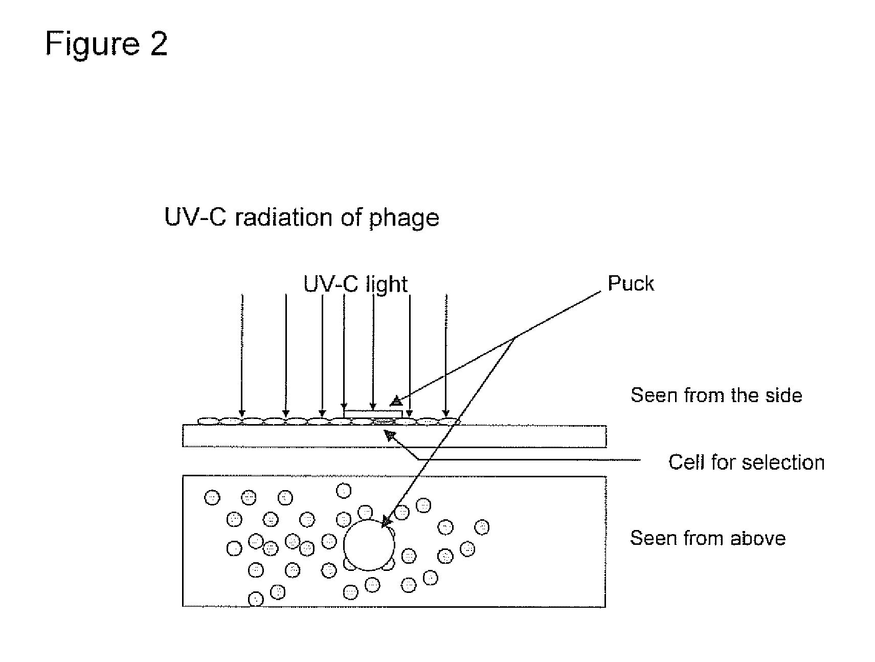
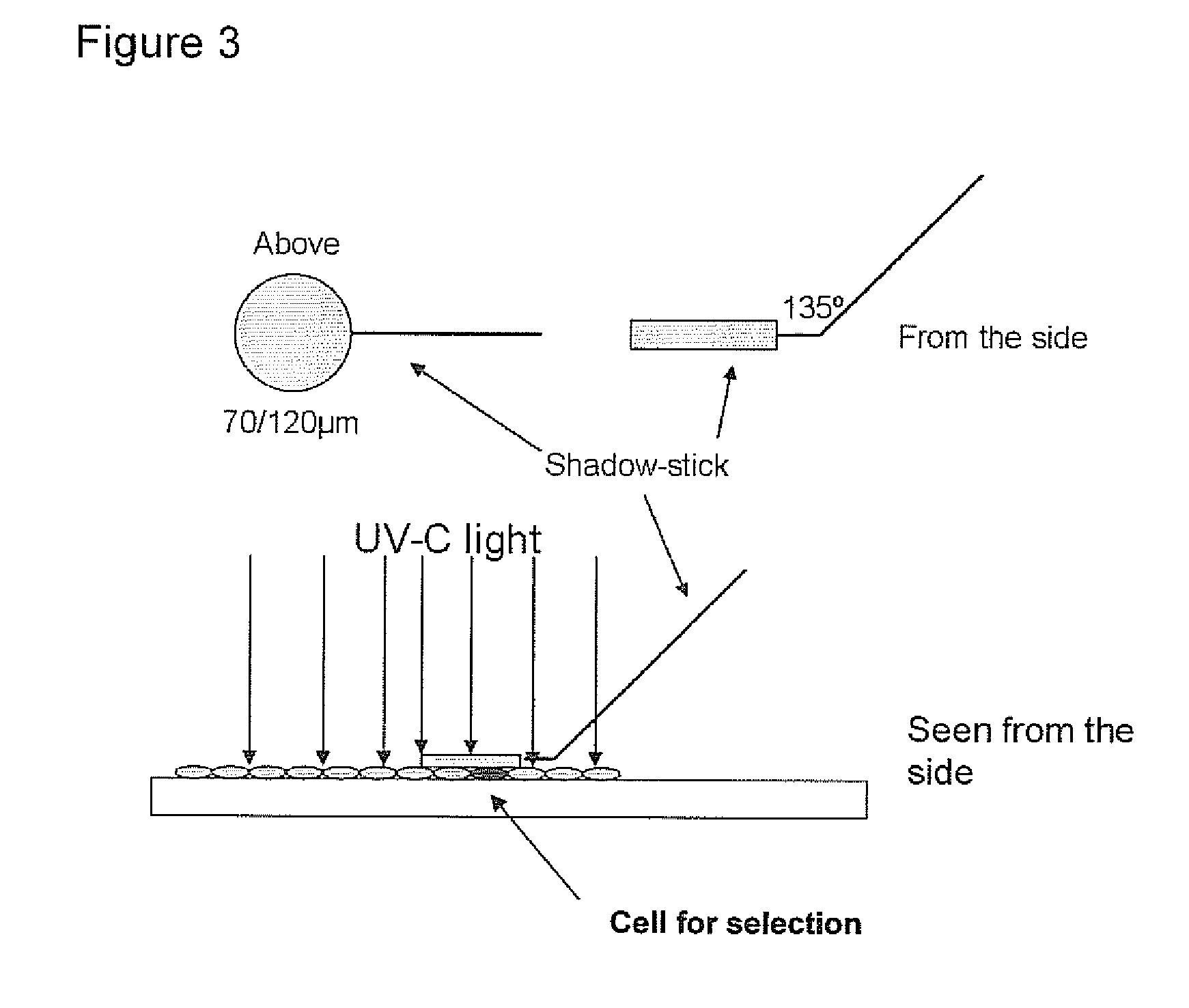
[0273]Foetal cell specific antibody fragments against cell markers are generated by use of phage display.

[0274]Based on the identification of foetal cells by FISH, the localization of the foetal cell on the slides is known. The identified foetal cell is subjected to biopanning whereby single chain fragment variable (scFv) antibody fragments are generated.

[0275]The selection of antibody fragments may be preformed as follows.[0276]1. The slide is blocked for 1 hour in 2% MPBS (skimmed milk powder in 1×PBS) at room temperature in order to reduce the amount of unspecific binding. This is done by subjecting the slide into MPBS in a holder for slides.[0277]2. The slide is washed one time in 1×PBS[0278]3. Biopanning: the scFv phage library (McCafferty et al. 1990) is incubated with the slide in 2% MPBS for one hour, with gentle shaking. A suitable library may have a semi-synthetic VH+VL repertoire and two single framework repertoires. An...


PUM

PropertyMeasurementUnit
temperatureaaaaaaaaaa

Abstract

The present application relates to methods for identification of foetal cells and generation and isolation of binding members recognising foetal cells. Said methods may further be used for other purposes relating to characterisation of biological samples and biological antigens. The methods are characterised by the applicability in situations where the interesting objects are present in a limited amount, or where the interesting objects are intermixed with other material, thus the methods are suitable for use in situations where the ratio of the interesting material compared to other material is low. The application discloses methods for use of detecting foetal cells and method of generating / isolating binding members towards antigenic material of low abundancy.

Description

REFERENCE TO RELATED APPLICATIONS[0001]This U.S. continuation patent application claims priority of U.S. patent application Ser. No. 12 / 096,594, filed Jun. 6, 2008, which is a U.S. National Phase of International Patent Application Serial No. PCT / DK2006 / 000693, filed Dec. 7, 2006, which claims priority of Denmark Application Serial No. PA 2005 01745, filed Dec. 8, 2005, all of which are incorporated herein by reference in their entirety. All patent and non-patent references cited in the application, or in the present application, are also hereby incorporated by reference in their entirety.FIELD OF INVENTION[0002]The present invention relates to methods for detection and characterisation of cells, tissue or target antigenic material of very limited availability. The analysis of rare cells, such as foetal cells in a maternal blood sample or a micometastatic cell, is complicated by the low abundance of such cells and because the biological sample often will comprise a majority of other...

Claims


Application Information

Patent Timeline
Patent Type & Authority Applications(United States)
IPC IPC(8): G01N33/569C07K16/28C07K14/00G01N21/64C12Q1/68G01N33/82C07K1/14C07K14/705
CPCC12N5/0081C12N15/1037G01N33/566C12Q1/6841C12Q1/6879C12Q1/6806C12Q2600/156
Inventor KOLVRAA, STEENCHRISTENSEN, BRITTASCHELDE, PALLESORENSEN, MORTEN DRAEBYKRISTENSEN, PETER
Owner AARHUS UNIV
Why Eureka
Try Eureka