
High-Throughput Platform Comprising Microtissues Perfused With Living Microvessels

Active Publication Date: 2012-04-05
RGT UNIV OF CALIFORNIA
View PDF2 Cites 15 Cited by
  • Summary
  • Abstract
  • Description
  • Claims
  • Application Information

AI Technical Summary

Benefits of technology

[0008]We have solved some of the above problems by mimicking the in-vivo formation of microvessels using a microfabricated platform combined with living cells and an extracellular matrix. Prior attempts have microfabricat

Problems solved by technology

Hence, the resulting channels are not true microvessels and do not mimic the dynamics of microvessel function.

Method used


Image

  • High-Throughput Platform Comprising Microtissues Perfused With Living Microvessels
  • High-Throughput Platform Comprising Microtissues Perfused With Living Microvessels
  • High-Throughput Platform Comprising Microtissues Perfused With Living Microvessels

Examples


Embodiment Construction

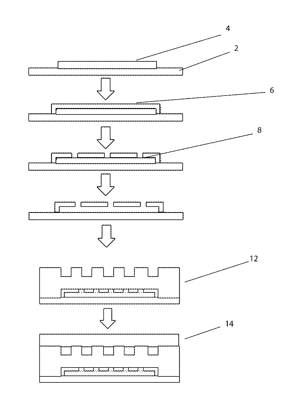
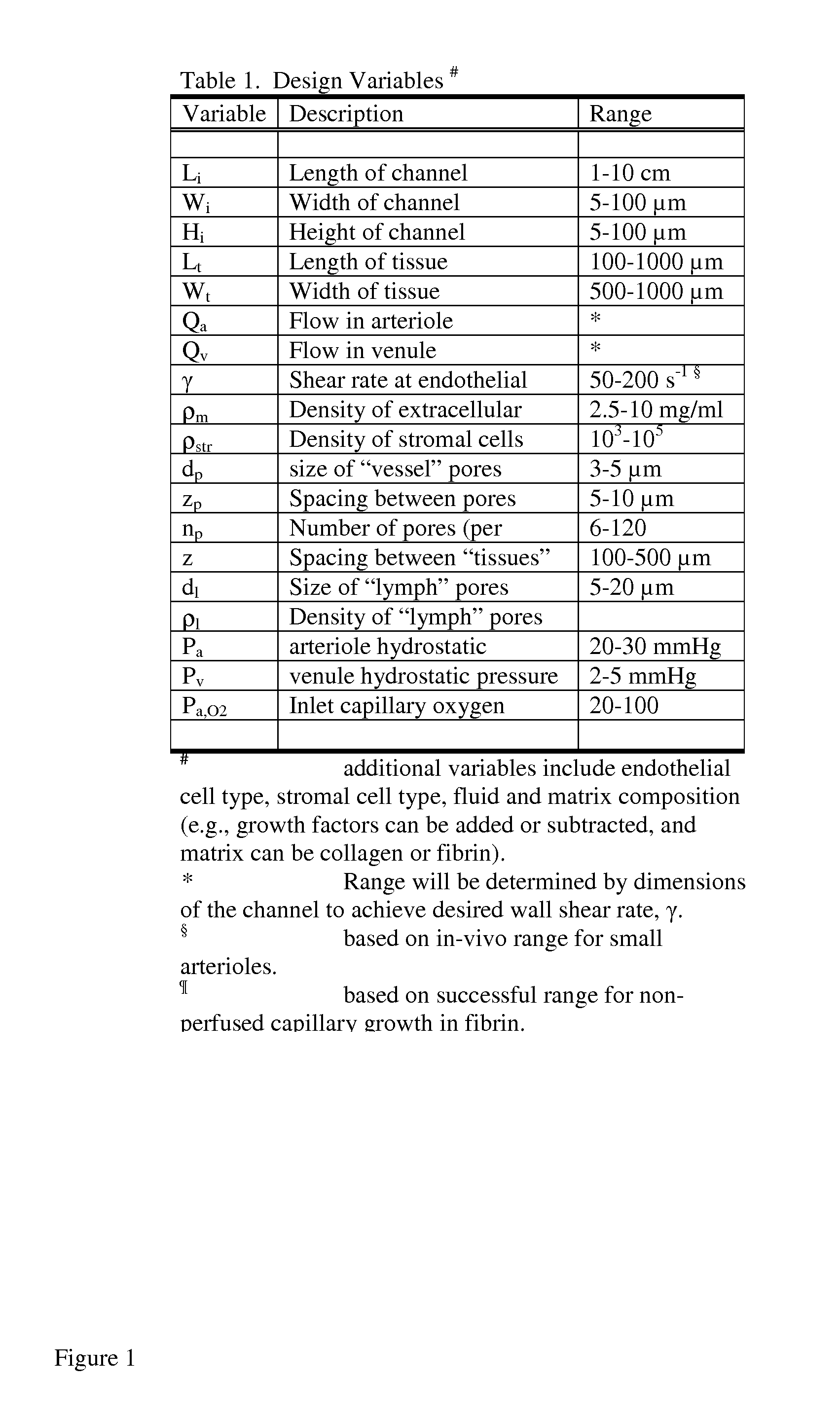
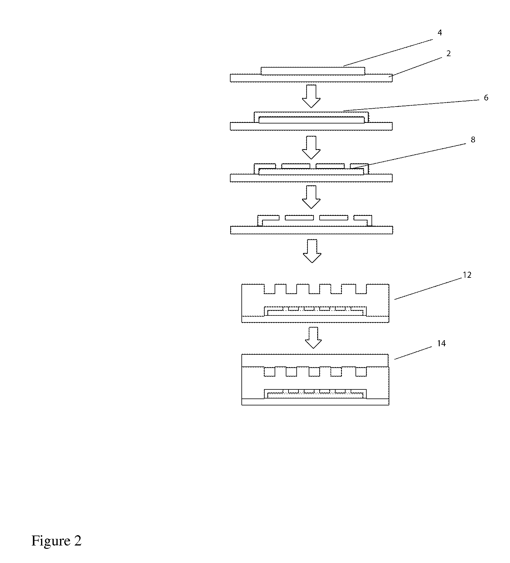
[0040]The term “microvessels” or “living microvessels” as used herein include arterioles, capillaries, venules, and lymphatics vessels. These living microvessels produced by the various embodiments connect the microfluidic channels to the microtissue. These microvessels are formed within the “pores” structures / channels located within the microfluidic channels (see below). They are metabolically active.

[0041]The term “microfluidic channels” as used here refer to the disclosed “arteriole” or “venuole” supplying channels, with respect to supplying or removing material from the microtissue compartment. “Arterioles” supply nutrients / fluid etc. to the microtissue; whilst “Venuoles” remove nutrients / fluid from the microtissue. These microfluidic channels are created by microfabrication technology and are not considered “3-D metabolically active” or lying vessels. (i.e. the above microvessels).

[0042]The term “microtissue compartment” as used here refers to a location where cells are grown. ...


PUM


Abstract

Provided is a process for creating a 3D metabolically active microtissue perfused with living microvessels which have a direct fluidic connection with neighboring microfluidic channels. The process comprises preparing a template comprising a plurality of channels, and creating a network within said channels, said network comprising microfluidic channels, metabolically active living microvessels, and microtissues. The microvessels can sprout from said microvessels and / or form within the microtissue in response to a stimulus applied from said microfluidic channels or stimulus derived from the said tissues. In another embodiment, a device is provided comprising a supportive structure, one or more microfluidic channels, one or more microtissue compartments, and one or more microvessels, whereby the microvessels connect said microfludic channels and microtissue and perfuse the microtissue to deliver fluid from the microfluidic channels to the microtissues.

Description

CROSS REFERENCE TO RELATED APPLICATIONS[0001]This application claims the benefit of Provisional Patent Application No. 61 / 404,619, filed on Oct. 5, 2010, which is incorporated by reference herein in its entirety.GOVERNMENT FUNDING / SUPPORT[0002]This invention was made with Government support with a grant from the National Institutes of Health (RC1-ES018361). The Government has certain rights in this invention.FIELD OF THE INVENTION[0003]The invention relates to the creation of in vitro microtissues; more particularly microtissues constructed on a microfabricated platform and perfused with living microvessels.BACKGROUND OF THE INVENTION[0004]The challenge to create the microenvironment enabling growth of an in-vitro microtissue perfused with living microvessels (e.g., arterioles, capillaries, and venules) represents a completely new paradigm in the creation of 3-D tissues. By definition, a 3-D tissue requires enhanced transport of nutrients and waste relative to 2-D monolayer cultures...

Claims


Application Information

Patent Timeline
IPC IPC(8): C40B30/06C40B40/02C40B50/06
CPCC12N5/0062C12N5/0691C12N2533/30G01N33/5082G01N33/5008G01N33/5011C12N2535/00
Inventor GEORGE, STEVEN C.HUGHES, CHRISTOPHER C.W.LEE, ABRAHAM P.MOYA, MONICAHSU, YU-HSIANG
Owner RGT UNIV OF CALIFORNIA
Learn More