Generating views of medical images

a technology of medical images and views, applied in the field of generating views of medical images, can solve the problems of time-consuming steps and manual generation of views, and achieve the effect of reducing the number of steps and reducing the cost of manual generation

Active Publication Date: 2012-04-26
KONINKLIJKE PHILIPS ELECTRONICS NV
View PDF11 Cites 16 Cited by
  • Summary
  • Abstract
  • Description
  • Claims
  • Application Information

AI Technical Summary

Benefits of technology

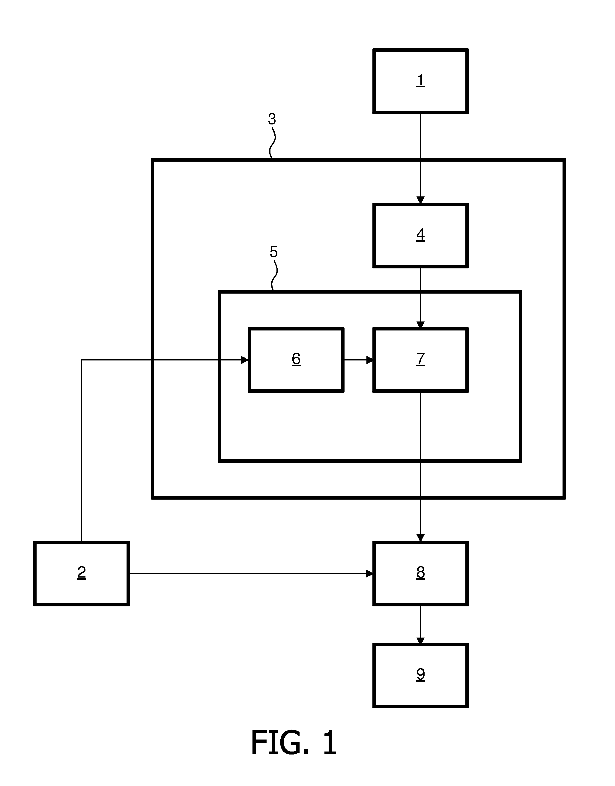
[0008]The system thus provides a view of the medical image which is associated, via the set of viewing parameters, with the suspected diagnosis. By automatically generating a view relevant to a suspected diagnosis, the burden of manually generating the view for confirming or rejecting that diagnosis is taken away from the clinician. Accordingly, the viewing of medical images for performing diagnoses becomes more efficient because fewer manual operations are needed. Moreover, the view generation may become more consistent because the views are provided automatically.
[0009]The mapper may comprise a database for mapping a suspected diagnosis into a set of generic viewing parameters, and a transformer for transforming the set of generic viewing parameters into a set of image-specific viewing parameters, based on the content of the medical image. In many cases, the desired view is only available relative to some aspect of the image content. The mapping table and the transformer make the viewing parameters image-specific, taking this image content into account. This allows providing more accurate views enabling a particular suspected diagnosis to be confirmed or rejected.
[0016]An image finding-indicating means may be provided for indicating an image finding to which the view of the medical image is directed. This way, the user of the system is prompted with the image finding he could make with this view. It allows the user to understand the reason why the system has generated this view. Moreover, it makes it easy for the user to know what he should look for in the image. A suspected diagnosis-indicating means may be provided for indicating to a user a suspected diagnosis associated with the view of the medical image. This helps to remind the radiologist of the suspected diagnosis.
[0017]The mapper may be arranged for generating an ordered list of sets of viewing parameters; the output may be arranged for providing respective views of the medical image in accordance with the respective sets of viewing parameters, in the order defined by the ordered list. This allows efficient reviewing of a sequence of useful images which are relevant to making a diagnosis.

Problems solved by technology

In this process, the manual generation of views can be very time-consuming.
These steps may be time-consuming.

Method used


Image

  • Generating views of medical images
  • Generating views of medical images
  • Generating views of medical images

Examples


Embodiment Construction

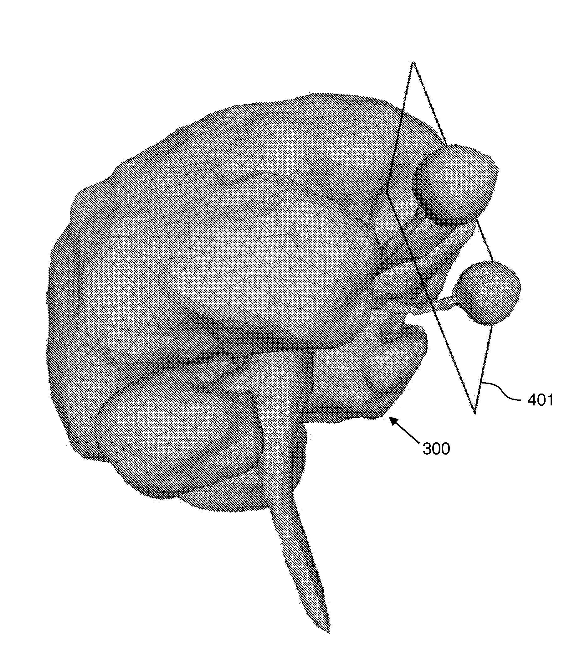
[0037]For a given patient, based on the patient's data, in particular acquired medical image data sets and one or more initial hypotheses of a disease or diagnosis, the radiologist has to confirm or reject the hypotheses to derive a differential diagnosis. To this end, he maps a hypothesis to hypothesis-specific image findings that would confirm or reject such a hypothesis. If the clinician is able to demonstrate such an image finding, he may confirm the hypothesis, otherwise he may reject the hypothesis. Alternatively, the capability to demonstrate the presence of particular image findings may be part of a process of diagnosing a patient. To confirm or reject the presence of an image finding, the clinician generates appropriate views of the patient's data, and identifies the findings in the views. The generation of views of the patient data to verify image findings supporting or rejecting a given hypothesis is a time-consuming, largely manual process performed by the radiologist / do...


PUM


Abstract

A system for generating a view of a medical image comprises an input (1) for receiving information indicative of a suspected diagnosis of a patient. An input (2) for receiving a medical image of the patient. A mapper (3) for mapping the suspected diagnosis of the patient to a set of viewing parameters for viewing the medical image. A view generator (8) for providing a view of the medical image in accordance with the set of viewing parameters. A database (4) for mapping a suspected diagnosis into a set of generic viewing parameters. A transformer (5) for transforming the set of generic viewing parameters into a set of image-specific viewing parameters based on content of the medical image.

Description

FIELD OF THE INVENTION[0001]The invention relates to generating a view of a medical image.BACKGROUND OF THE INVENTION[0002]As medical imaging technologies advance, more and more image data sets are acquired by medical scanners to facilitate the diagnosis. For a given patient, based on information collected about the patient, the clinician has the task of establishing a diagnosis of the patient. To this end, the clinician may have a hypothetical diagnosis he wishes to confirm or reject, based on results of patient examination, such as medical images or laboratory results. In the case of medical images, the clinician maps the hypothesis into image findings which can help in confirming or rejecting the diagnosis. To obtain the image findings, the clinician not only needs to acquire the necessary medical images, using an imaging modality such as x-ray, CT, or MR, but he also needs to generate appropriate views of the acquired medical image data. Such views may be generated using an imag...

Claims


Application Information

Patent Timeline
Patent Type & Authority Applications(United States)
IPC IPC(8): G06T1/00
CPCG06T2210/41G06T19/00
Inventor LEHMANN, HELKOWEESE, JUERGENBOUGHORBEL, SABRIQIAN, YUECHENSEVENSTER, MERLIJNSILFEN, ERIC ZACHERY
Owner KONINKLIJKE PHILIPS ELECTRONICS NV
Why Eureka
Try Eureka