Medical image diagnosis assisting apparatus, method, and program

a technology of medical image and diagnostic equipment, applied in the field of medical image diagnosis assisting technology, can solve problems such as cardiac arrest and inability to recover

Active Publication Date: 2012-04-26
FUJIFILM CORP
View PDF1 Cites 21 Cited by
  • Summary
  • Abstract
  • Description
  • Claims
  • Application Information

AI Technical Summary

Benefits of technology

[0026]According to the present invention, a superimposed image representing the cardiac function evaluation value and myocardial infarction rate in a superimposing manner such that they are distinguishable from each other in a coordinate system capable of representing each position of the heart in the three-dimensional medical image may be outputted. This allows calculation results of the cardiac function evaluation value and myocardial infarction rate to be integrally visualized, thereby clearly indicating whether or not a myocardial infarction is developed in a portion where cardiac function is deteriorated. Consequently, distinction between angina pectoris and myocardial infarction may be made more easily and accurately at the time of image diagnosis.

Problems solved by technology

In most cardiac diseases, the supply of blood flow to the myocardium is stopped and the myocardium becomes unable to work, thereby causing cardiac arrest.
Whereas in the case of angina pectoris, the myocardium can be recovered, it cannot be recovered in the case of myocardial infarction because the myocardium has already been necrotized.

Method used


Image

  • Medical image diagnosis assisting apparatus, method, and program
  • Medical image diagnosis assisting apparatus, method, and program
  • Medical image diagnosis assisting apparatus, method, and program

Examples


Embodiment Construction

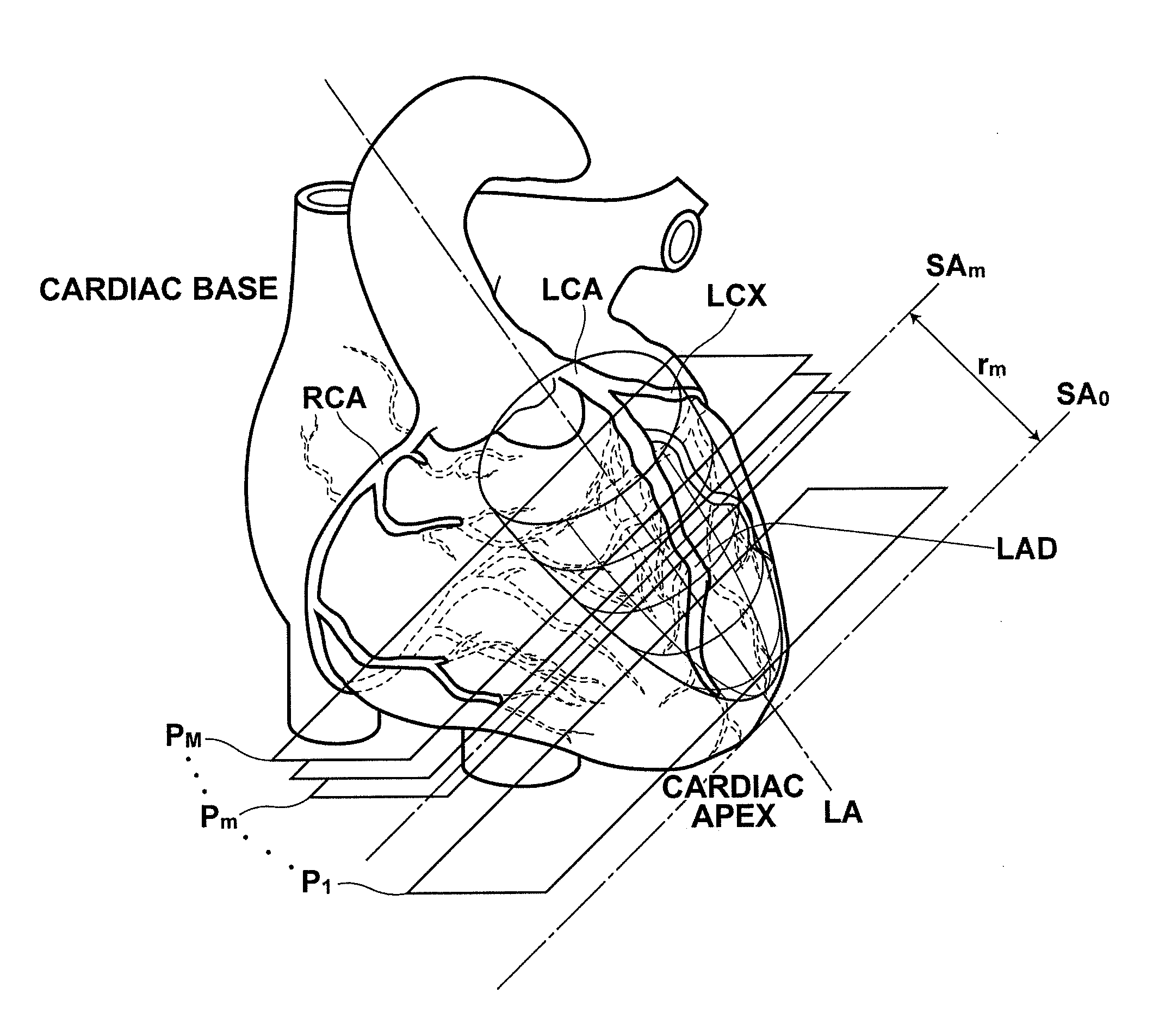
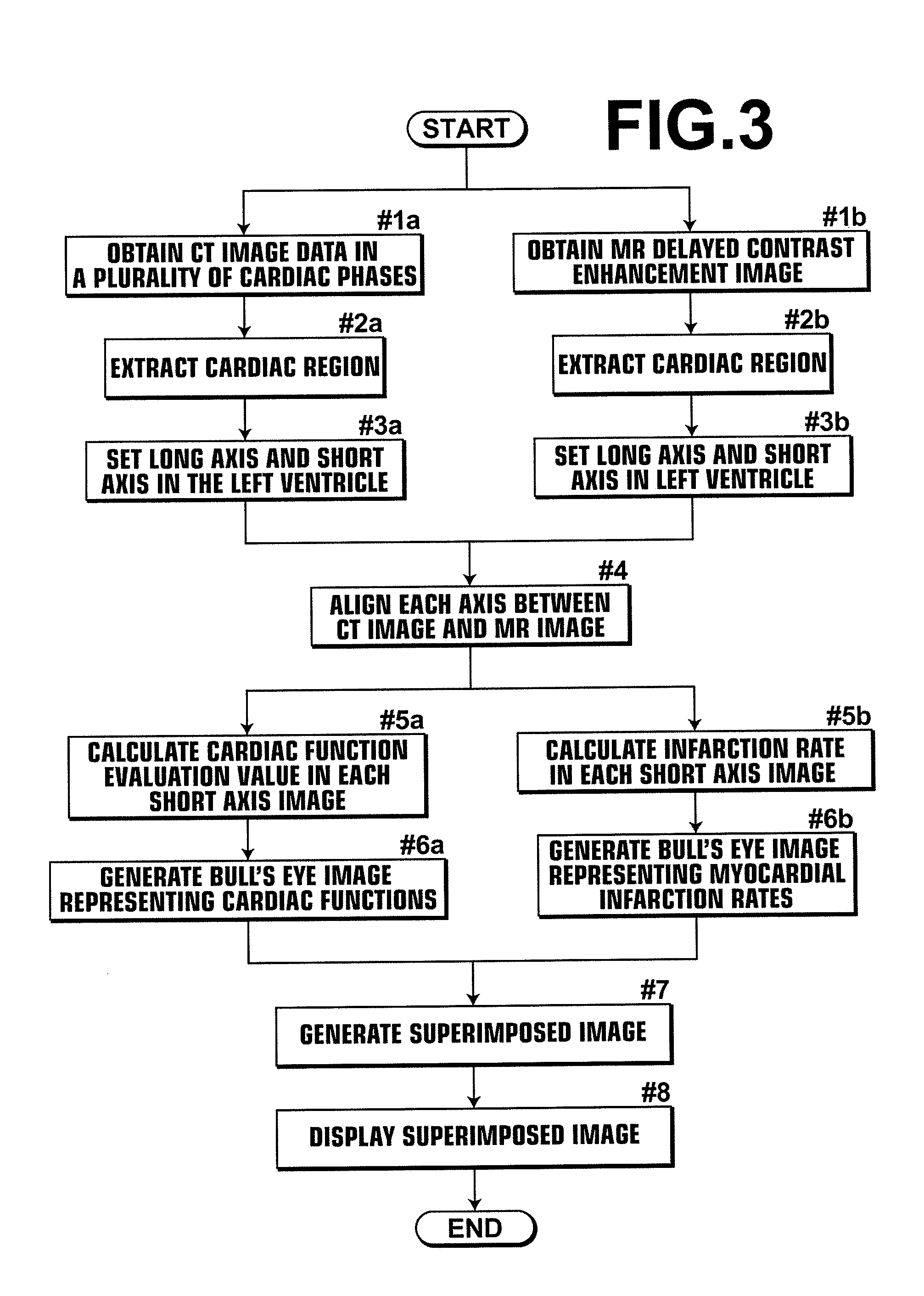
[0042]Hereinafter, a medical image diagnosis system that employs a medical image diagnosis assisting apparatus of an embodiment of the present invention will be described, taking cardiac analysis processing, as an example, in which cardiac function analysis results based on three-dimensional medical images in a plurality of different cardiac phases obtained by multi-detector-row computed tomography and analysis results of infarction areas of myocardium based on a three-dimensional medical image obtained by MR delayed contrast enhancement are displayed in a superimposing manner.

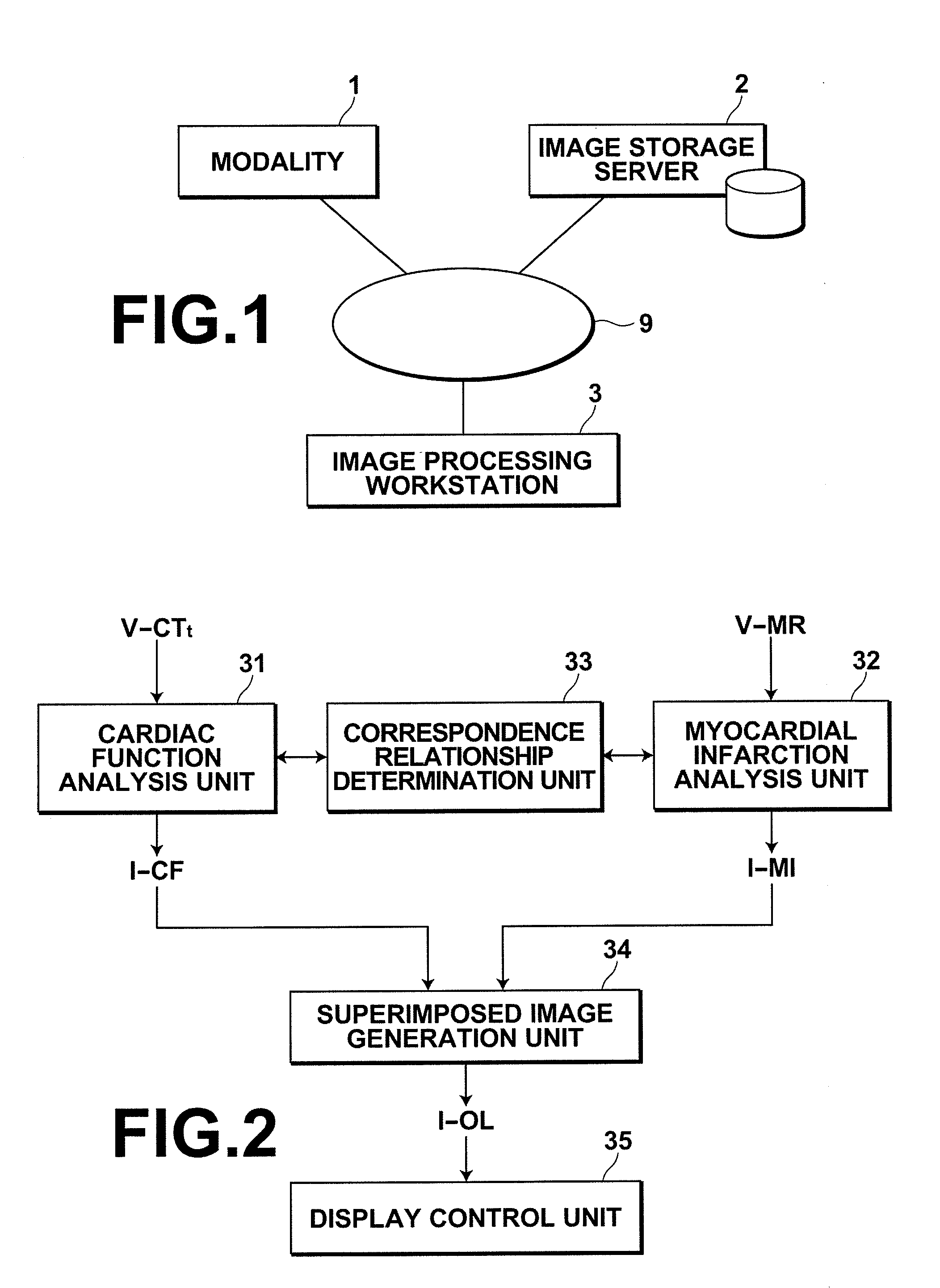
[0043]FIG. 1 is a hardware configuration diagram of the medical image diagnosis system, illustrating an overview thereof. As illustrated in FIG. 1, the system includes modality 1, image storage server 2, and image processing workstation 3 communicatively linked to each other via network 9.

[0044]Modality 1 is an apparatus for imaging a chest region (cardiac region) of a subject to generate image data of a three...


PUM


Abstract

Using a three-dimensional medical image representing a heart as input, a cardiac function analysis unit calculates a cardiac function evaluation value representing a cardiac function with respect to each of predetermined portions of the heart, using a three-dimensional medical image representing the heart as input, a myocardial infarction analysis unit calculates a myocardial infarction rate representing a degree of myocardial infarction with respect to each of predetermined portions of the heart, and a superimposed image output unit outputs a superimposed image representing the cardiac function evaluation value and the myocardial infarction rate in a superimposing manner such that they are distinguishable from each other in a coordinate system capable of representing each position of the heart in the three-dimensional medical image.

Description

BACKGROUND OF THE INVENTION[0001]1. Field of the Invention[0002]The present invention relates to a medical image diagnosis assisting technology for outputting an image useful for image diagnosis of a cardiac region based on a three-dimensional medical image.[0003]2. Description of the Related Art[0004]Medical image analysis techniques have been advancing rapidly with a wide spread use of multi-detector row computed tomography in clinical practice. For example, in the field of cardiac medicine, it has become possible to obtain a plurality of three-dimensional images within one heartbeat. Along with this, various image diagnosis assisting methods using a three-dimensional medical image of cardiac region have been proposed.[0005]In most cardiac diseases, the supply of blood flow to the myocardium is stopped and the myocardium becomes unable to work, thereby causing cardiac arrest. Therefore, it is necessary to discover a myocardial abnormality by image diagnosis and determine the treat...

Claims


Application Information

Patent Timeline
Patent Type & Authority Applications(United States)
IPC IPC(8): A61B5/055
CPCA61B6/03A61B6/466A61B6/503A61B6/5247G06T2207/30048G06T2219/016G06T2200/24G06T2207/10081G06T2207/10096G06T19/20G06T2219/2012G06T7/0012
Inventor MASUMOTO, JUN
Owner FUJIFILM CORP
Why Eureka
Try Eureka