Region of interest determination for x-ray imaging

a x-ray imaging and region determination technology, applied in the field of x-ray imaging systems, can solve the problems of difficult to align body parts with difficult to align smaller body parts with the ion chamber, and difficult to align body parts with the fixed locations on the system

Active Publication Date: 2012-05-24
GENERAL ELECTRIC CO
View PDF4 Cites 26 Cited by
  • Summary
  • Abstract
  • Description
  • Claims
  • Application Information

AI Technical Summary

Problems solved by technology

Problems arise, however, in situations where it is difficult to align body parts with the fixed locations on the system, especially when these fixed locations are not properly adapted to the patient anatomies, patient sizes, and so forth.
By way of example, pediatric imaging is especially challenging because it is often difficult to align smaller body parts with the ion chambers of the imaging system.
Because the exposure measurement devices, such as ion chambers serve as integrators of received radiation, misalignment may result in under or over estimating the actual radiation actually applied to the anatomy of interest.

Method used


Image

  • Region of interest determination for x-ray imaging
  • Region of interest determination for x-ray imaging
  • Region of interest determination for x-ray imaging

Examples


Embodiment Construction

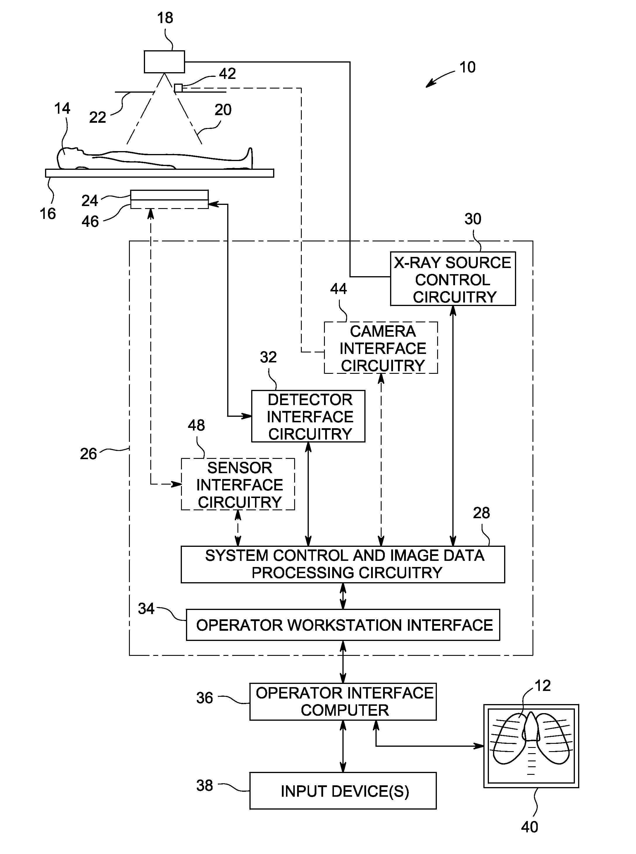
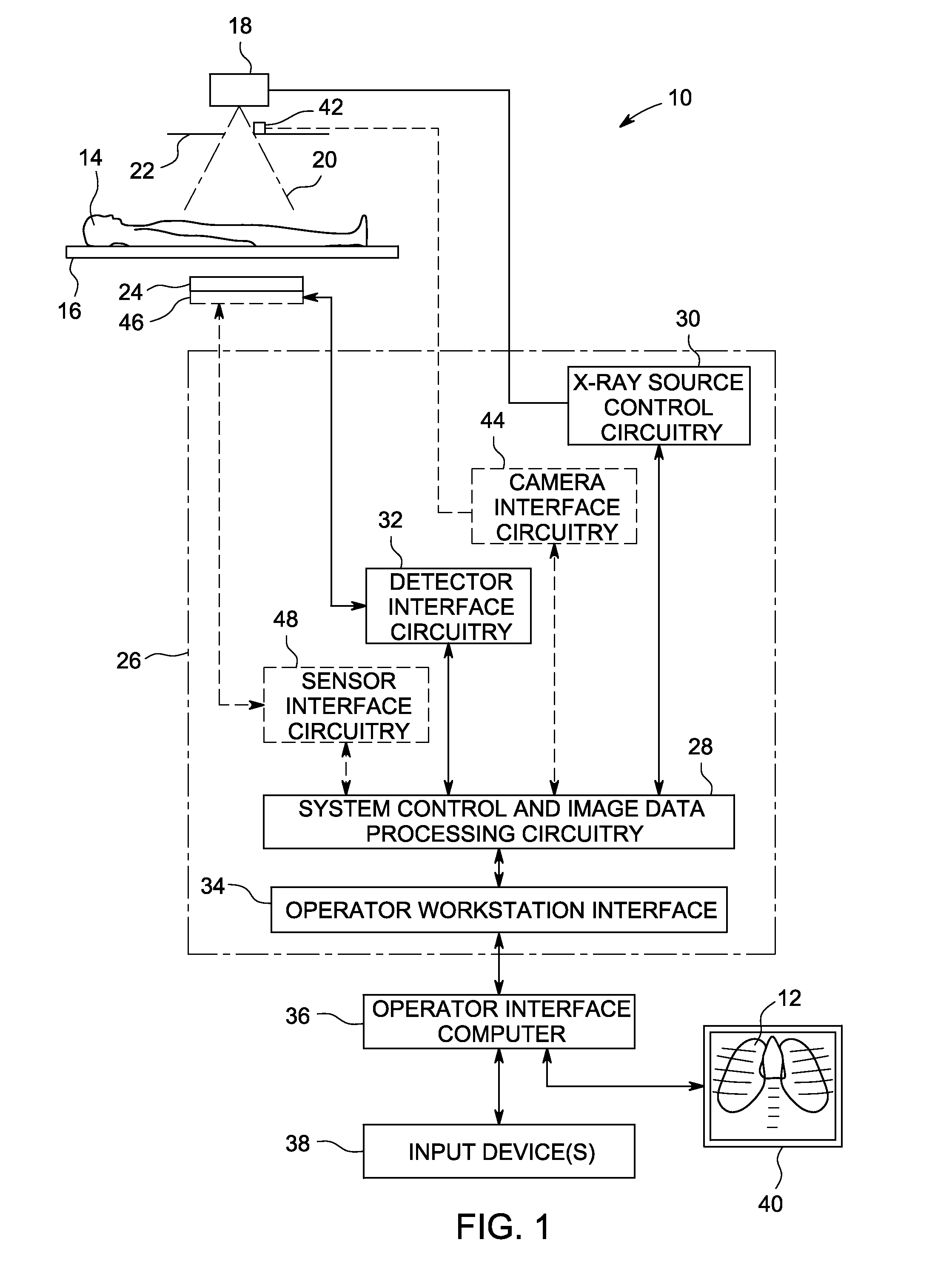
[0017]Referring to FIG. 1, an X-ray imaging system 10 is illustrated that allows for identification of a region of interest and exposure control based upon the region of interest. The X-ray imaging system 10 is adapted for generating images 12 of a subject 14. In a medical diagnostic context, the subject 14 may be positioned on a support 16. An X-ray source 18 is adapted to produce a beam of radiation 20 which passes through collimator 22. The radiation traverses the subject, with some of the radiation being attenuated or absorbed, and resulting radiation impacting a detector 24.

[0018]A control and processing system 26 is coupled to both the radiation source and the detector 24. In general, this system allows for regulation of operation of both the source and the detector, and permits collection of information from the detector for reconstruction of useful images. In the illustrated embodiment, for example, the control and processing system 26 includes system control and image proce...


PUM

PropertyMeasurementUnit
computed tomographyaaaaaaaaaa
sizesaaaaaaaaaa
field of viewaaaaaaaaaa

Abstract

A radiography system allow for user determination of a region of interest on a subject prior to X-ray exposure. The region of interest is defined by user interaction with an image, a pointer system, or the like. The region of interest is then translated to the imaging coordinate system, such as in the plane of a digital detector. The region is then used for exposure control during an imaging sequence, either in an open or closed-loop manner.

Description

BACKGROUND OF THE INVENTION[0001]The present invention relates generally to field of X-ray imaging systems, and more particularly to optimization to radiation dosage by specific definition of a region of interest to be imaged.[0002]Radiography systems, such as X-ray systems, are used to generate images showing internal features of a subject. In the medical context, such systems are used for viewing internal anatomies and tissues, such as for diagnostic purposes. In modern projection X-ray systems, for example, X-rays are generated by an X-ray source and are directed to a patient or other subject. The X-rays transfer through the subject, and are absorbed or attenuated by internal features. The resulting X-rays impact a digital detector where image data is generated. Collecting the image data allows for reconstruction of a useful image. Similar techniques are used for computed tomography, fluoroscopy and tomosynthesis image generation.[0003]It is a general goal in radiography to acqui...

Claims


Application Information

Patent Timeline
Patent Type & Authority Applications(United States)
IPC IPC(8): G01N23/04
CPCA61B6/469A61B6/542A61B6/488
Inventor JABRI, KADRI NIZARWALIMBE, VIVEKSAUNDERS, ROWLANDARESTE, ROMAIN
Owner GENERAL ELECTRIC CO
Why Eureka
Try Eureka