Apparatus and Method for Cutting Costal Cartilage

a costal cartilage and apparatus technology, applied in the field of medical equipment, can solve the problems of reducing the skill and time required to fashion cartilage slices, increasing uniformity, etc., and achieve the effects of reducing skill and time, increasing the uniformity of cut specimens, and increasing uniformity

Active Publication Date: 2012-07-26
RGT UNIV OF CALIFORNIA
View PDF3 Cites 15 Cited by
  • Summary
  • Abstract
  • Description
  • Claims
  • Application Information

AI Technical Summary

Benefits of technology

[0011]A double-bladed cutting device providing a practical method for obtaining costal cartilage specimens in both an operating room and research setting is disclosed. The device reduces the skill and time required to fashion cartilage slices, while increasing the uniformity of the cut specimens. Furthermore,

Problems solved by technology

The device reduces the skill and time required to fashion cartil

Method used


Image

  • Apparatus and Method for Cutting Costal Cartilage
  • Apparatus and Method for Cutting Costal Cartilage
  • Apparatus and Method for Cutting Costal Cartilage

Examples


Embodiment Construction

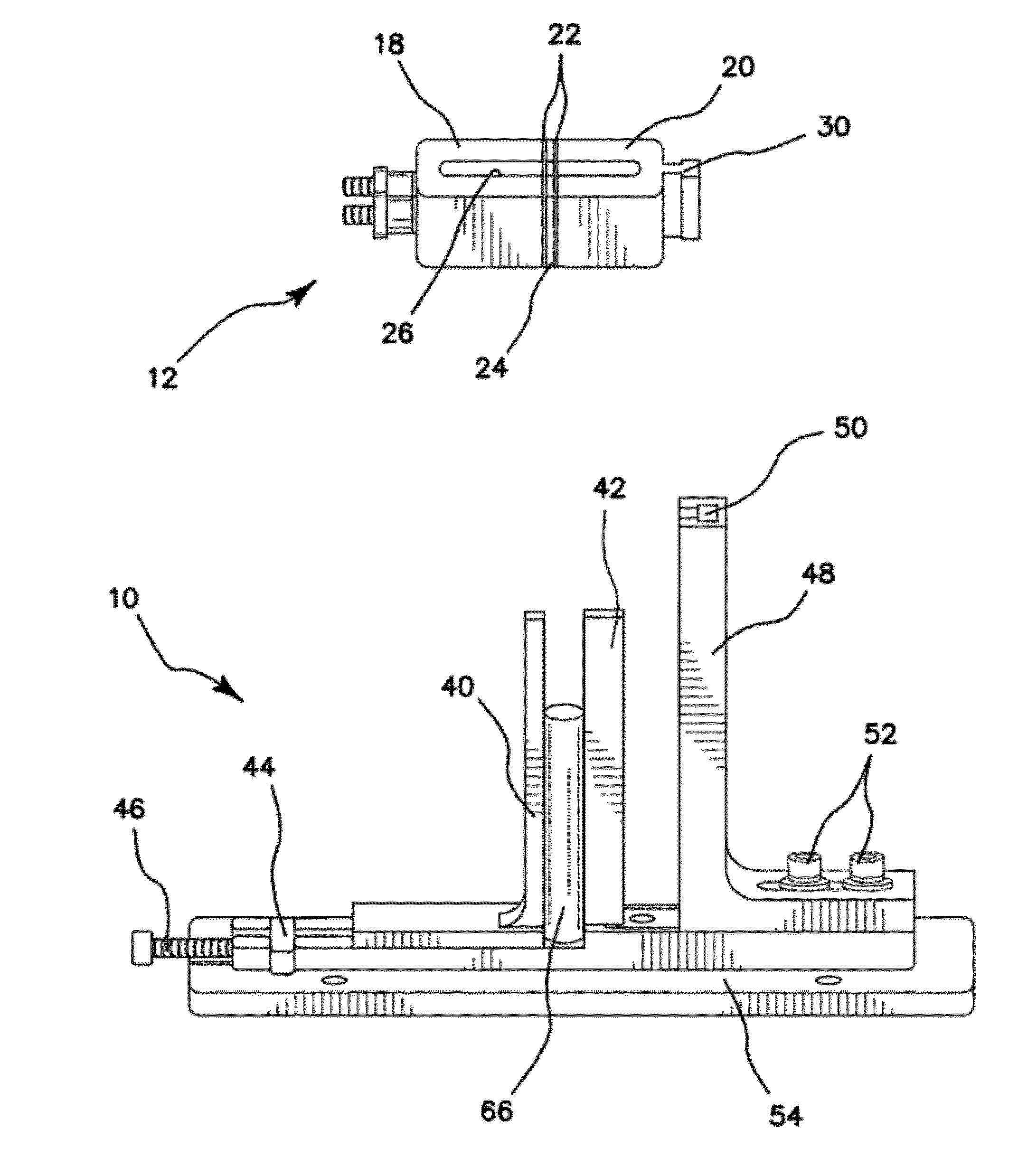
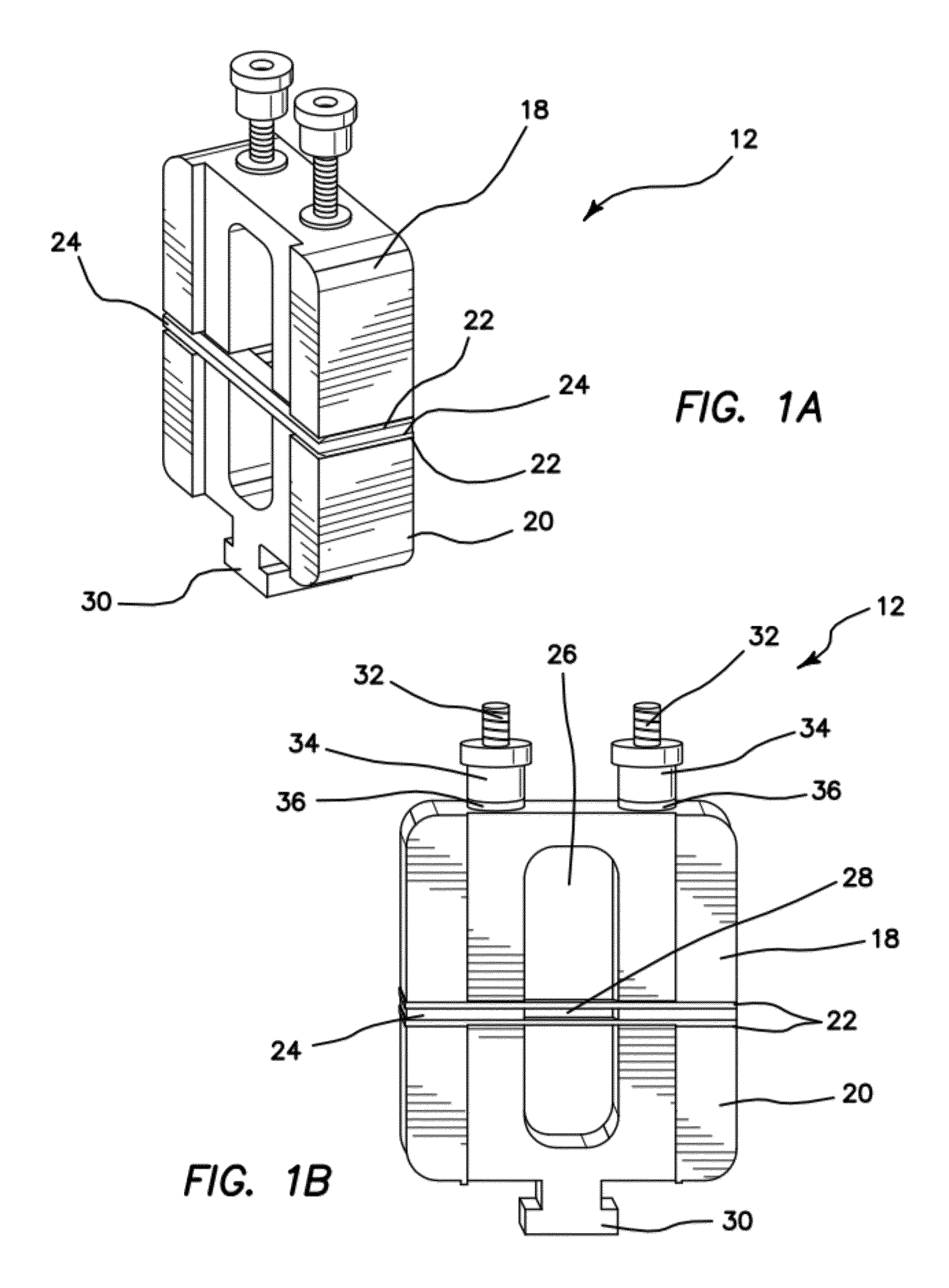
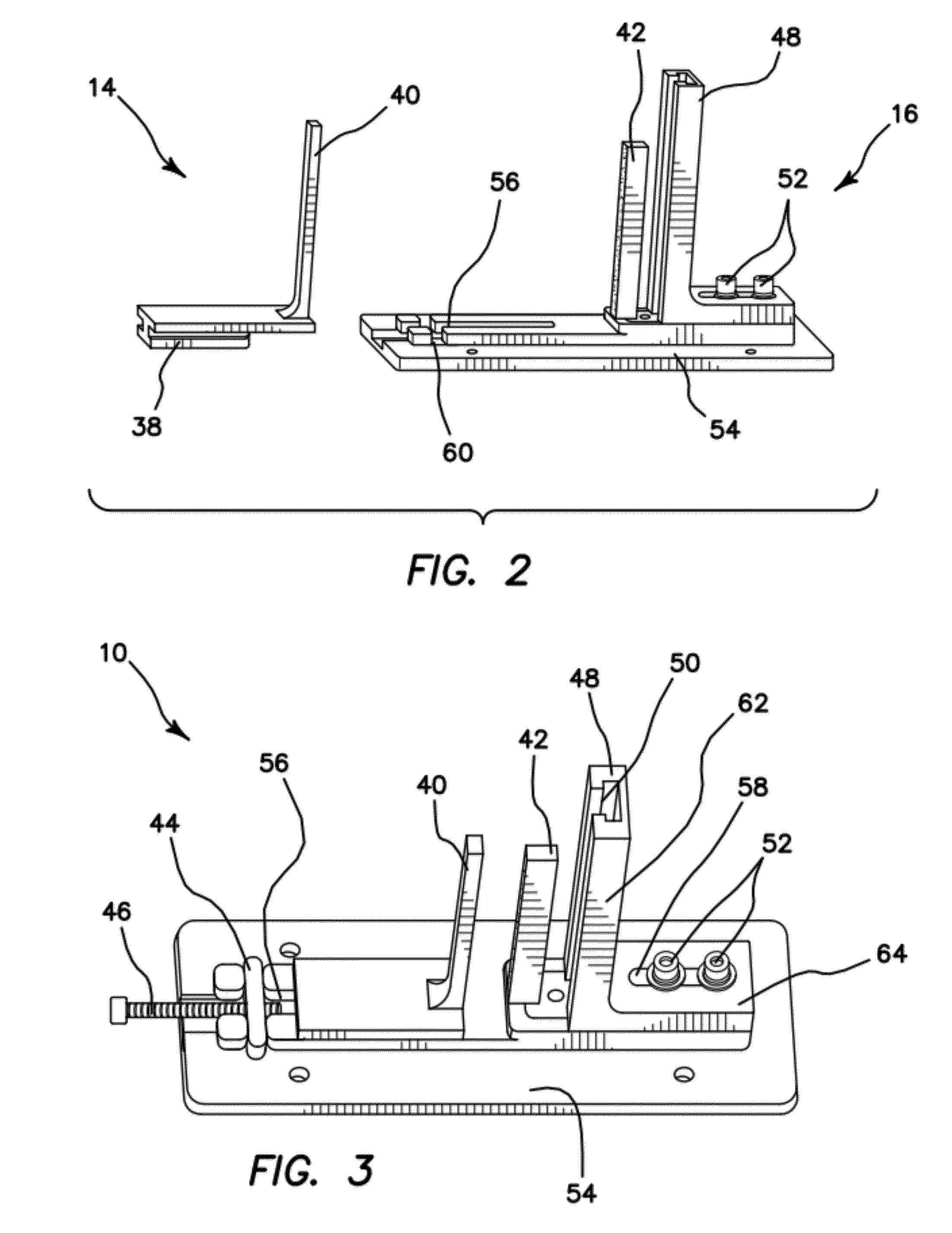
[0029]The current device comprises three main components, a cutting jig 12 seen in FIGS. 1A and 1B, a slide member 14 and a base 16 seen in FIGS. 2 and 3. When the slide member 14 and base 16 are coupled together as seen in FIG. 3 and as further described below, a specimen assembly 10 is formed.

[0030]Turning now to the cutting jig 12 and FIGS. 1A and 1B, it can be seen that the cutting jig 12 comprises two substantially U-shaped halves, namely a top half 18 and a bottom half 20. The bottom half 20 further comprises a substantially “T” shaped jig key 30 disposed along one of its outer edges. The key 30 helps to guide the cutting jig 12 through the specimen assembly 10 as is further detailed below. Disposed between the top half 18 and the bottom half 20 is a pair of parallel blades 22 separated by a pair of identical aluminum spacers 24. The halves 18, 20 effectively sandwich the pair of blades 22 and spacers 24 there between and due to their substantial U-shape, define an aperture 26...


PUM


Abstract

A double-bladed cutting device providing a practical method for obtaining costal cartilage specimens in both an operating room and research setting. The device reduces the skill and time required to fashion cartilage slices, while increasing the uniformity of the cut specimens. Furthermore, via an adjustable guide, slices can be obtained precisely from the central core of the rib. Although specimen lengths up to 4 cm are preferred, longer lengths can be attained without modification of the device. The cartilage specimen may be held in place within the device via means of compression or tension.

Description

STATEMENT OF GOVERNMENT SUPPORT[0001]This invention was made with government support under grant number DR090349 awarded by Department of Defense Deployment Related Medical Research Program and grant number 1R21DE019026 awarded by the National Institute of Dental and Craniofacial Research. The government has certain rights in the invention.BACKGROUND OF THE INVENTION[0002]1. Field of the Invention[0003]The invention relates to the field of medical tools, specifically to a cutting device used to obtain uniformly thick slices of cartilage from a costal (rib) cartilage sample.[0004]2. Description of the Prior Art[0005]Costal cartilage is becoming increasingly popular as a graft source for facial reconstruction. However, carving methods have not changed in decades, and continue to primarily rely upon detailed maneuvers with a scalpel. There are currently very few mechanical devices for shaping costal cartilage and the accuracy and precision of manual methods leave much to be desired.[00...

Claims


Application Information

Patent Timeline
IPC IPC(8): A61B17/56
CPCA61B17/1635A61B17/1693A61B17/1688A61B17/1606A61B17/1679A61F2/4644
Inventor WONG, BRIANMANUEL, CYRUSFOULAD, ALLEN
Owner RGT UNIV OF CALIFORNIA
Why Eureka
Try Eureka