Device and method for lasering biological tissue

a biological tissue and laser technology, applied in boring tools, dental tools, radiation therapy, etc., can solve the problems of no “smart” device for simultaneous and objective detection of pathological structures, unavoidable pain, and thermomechanical damage, so as to achieve maximum medical and biological compatibility, avoid undesirable collateral effects, and achieve more biological-medical compatibility

Inactive Publication Date: 2012-08-09
LUMERA LASER +1
View PDF9 Cites 5 Cited by
  • Summary
  • Abstract
  • Description
  • Claims
  • Application Information

AI Technical Summary

Benefits of technology

[0017]The embodiments may also be implemented to realize that that laser pulses having a temporal full width at half maximum in the picosecond range can now be used for advantageous effects. This range may provide the ablation efficiency, and the biomedical compatibility can also be optimized due to the optical depth of penetration. Accordingly, the thermal and mechanical stress may be limited.
[0018]The embodiments may also be implemented to realize that a marker that may render sites to be lasered or ablated visible for diagnosis can now be implemented simultaneously to the lasering. The marker may be a photosensitizer and / or can be activated by a laser beam or an LED continuously or pulsed with a suitable wavelength, duration and intensity.
[0019]The embodiments may also be implemented to realize that a site of the tissue to be lasered or ablated may be encapsulated by integrating an aspiration system in a laser beam decoupler / outcoupler.
[0020]The embodiments may be employed for abrading or ablating dentin, particularly when carious. Here, the application may utilize that carious dentin has a porous structure due to bacterial activity. The photosensitizer may gain access through this porous structure in embedding in the carious dentin to be ablated rather than applying to the surface of tissue material to be ablated.
[0021]When lasering biological tissue with a short-pulse laser such as a picosecond (ps) or femtosecond (fs) laser, microplasma may be generated within a thin surface layer at the focal position of the laser beam. Here, the microplasma may be ablated in a matter of nanoseconds or microseconds thus the biological tissue may not be ionized by interaction of the laser photons with the quasi-free electrons but minimally invasive thermo-mechanically fragmented. One general intention is to always generate the microplasma in the threshold region, i.e. always below the critical electron density (for the laser wavelength of 1064 nm: 1.03×10²¹ electrons / cm3) so that ablation with maximized medical and biological compatibility may be performed to avoid undesirable collateral effects. Especially, plasma temperatures greater than or equal to 5800 K (surface temperature of the sun) resulting in UV radiation and multiphoton ionization are to be avoided so that water molecules in the tissue are not ionized. In accordance with the present disclosure, an indirect energy input by photosensitizers injection and the usage of picosecond laser pulses provide more biological-medical compatibility. Especially, regarding the stress relaxation, an optical depth of penetration may result in no shock waves and enable treatments to be implemented painlessly.
[0022]In general, a surface site of the biological tissue to be treated may be scanned by the laser beam. Where this is concerned, the laser beam may have a top hat profile so that each sub-site focused by the laser beam is scanned with precisely one laser pulse. However, whether a top hat profile is provided or not, it is just as possible to achieve this by defining scanning each adjoining sub-sites with a single laser pulse with an overlap having a surface area smaller than half or smaller than some other fraction of the surface area of a sub-site. This may make it possible when the “cross-section of the laser beam” has a Gauβian profile that a sub-site substantially focused by the laser beam is pulsed substantially by a single laser pulse.

Problems solved by technology

In a practice treating oral tissue structures, the conventional “drill” still remains the main choice in dentistry because of its universality and low investment costs although it potential causes considerable thermo-mechanical damage (frictional heat, cracks, shock waves) coupled with the resulting unavoidable pain.
However, there is still no “smart” device for simultaneous and objective detection of pathological structures (e.g., caries) and therapy (e.g., cavitation preparation) with AUTO self-limiting stop for maximum bio-safety.
However, in many cases, undesirable thermal or other collateral effects were observed, or the ablation efficiency was inadequate.
In addition, none of these systems is capable of performing bio-safe detection and therapy.
However, particularly when more than three photons are involved, the risk was that such a high pulse energy involving very short laser pulses where the values attained as to power or intensity in the maximum pulse, harmful collateral effects may be materialized due to non-linear processes such as multi-photon ionization.
Accordingly, the thermal and mechanical stress may be limited.

Method used


Image

  • Device and method for lasering biological tissue
  • Device and method for lasering biological tissue
  • Device and method for lasering biological tissue

Examples


Embodiment Construction

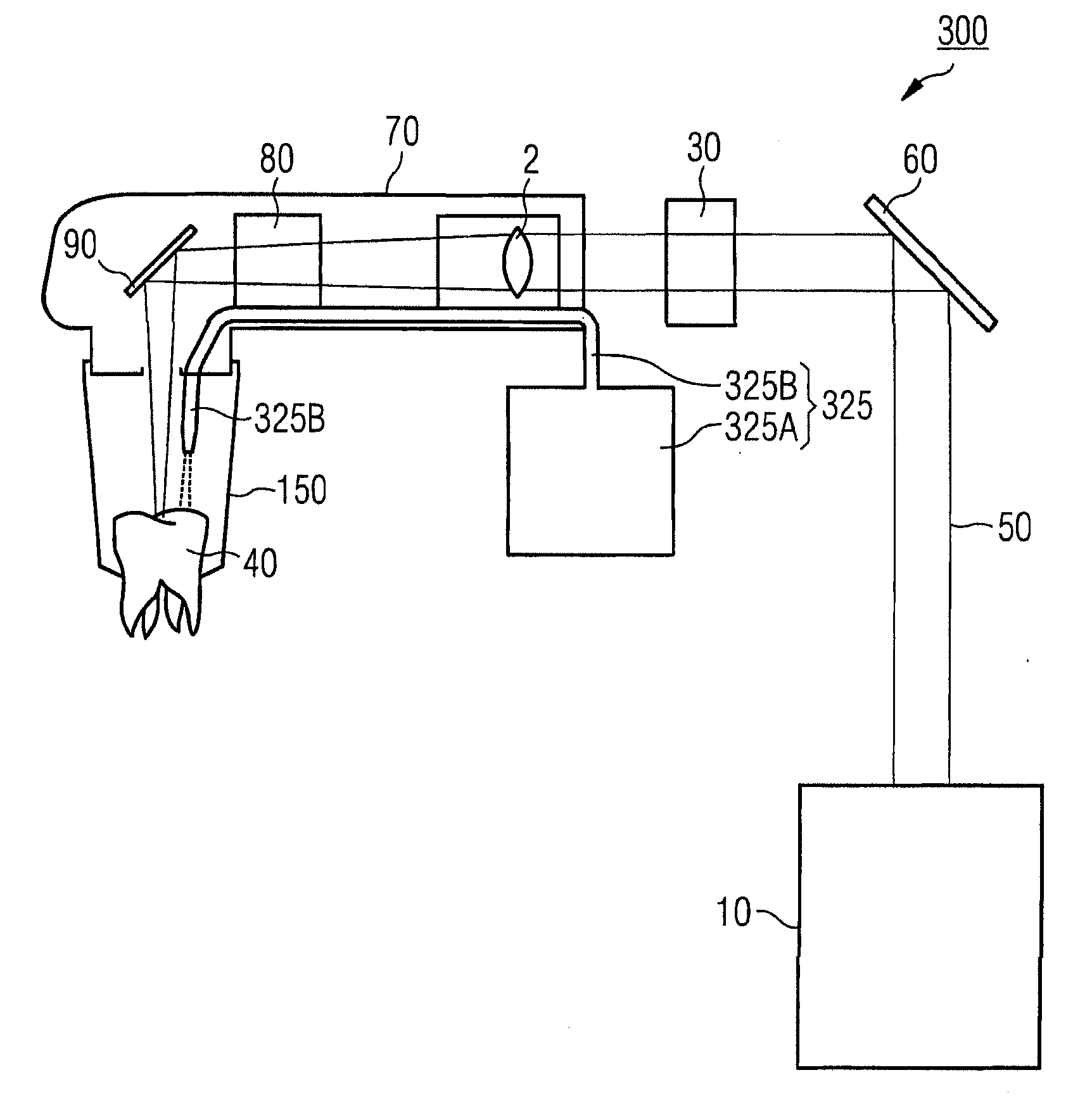
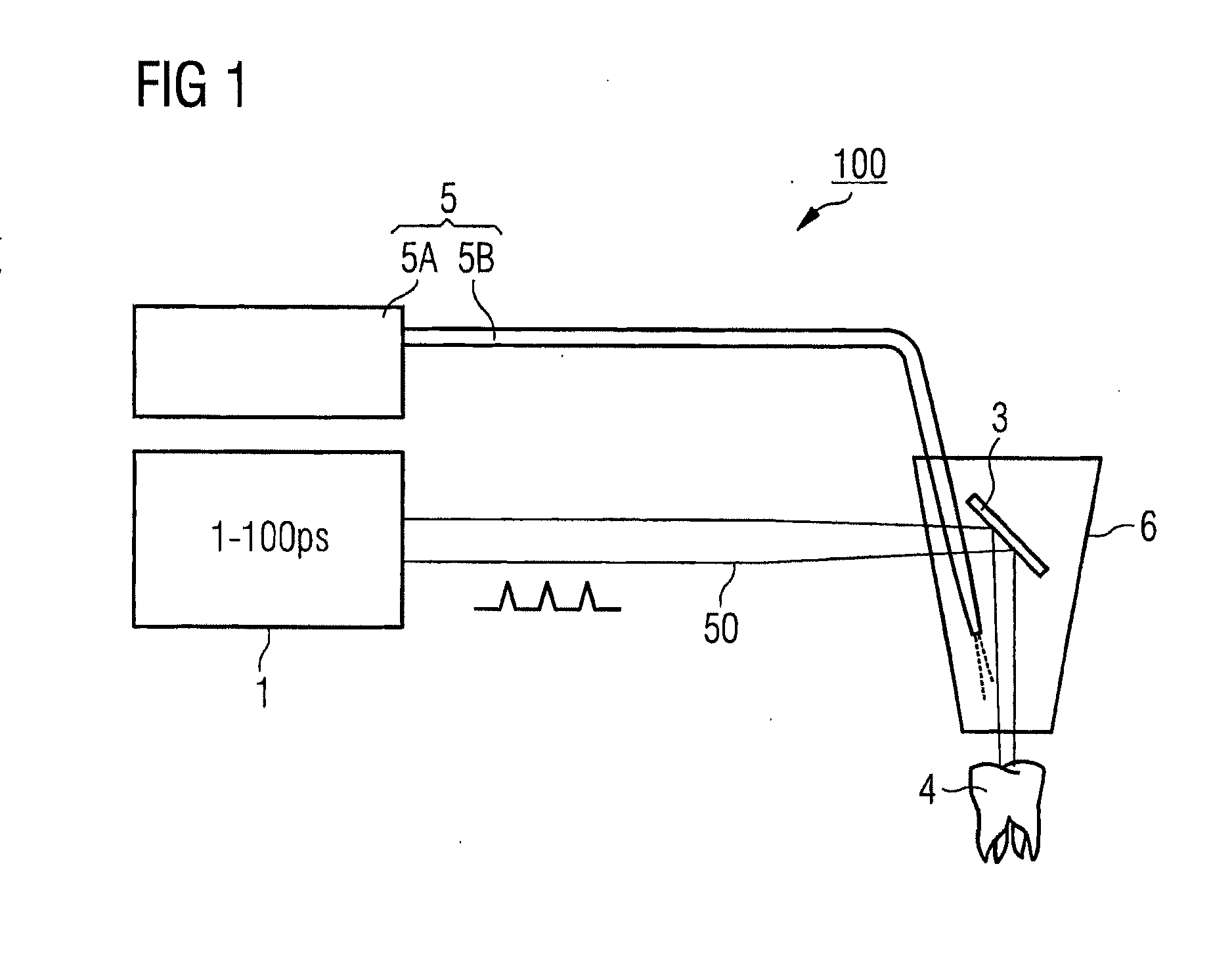
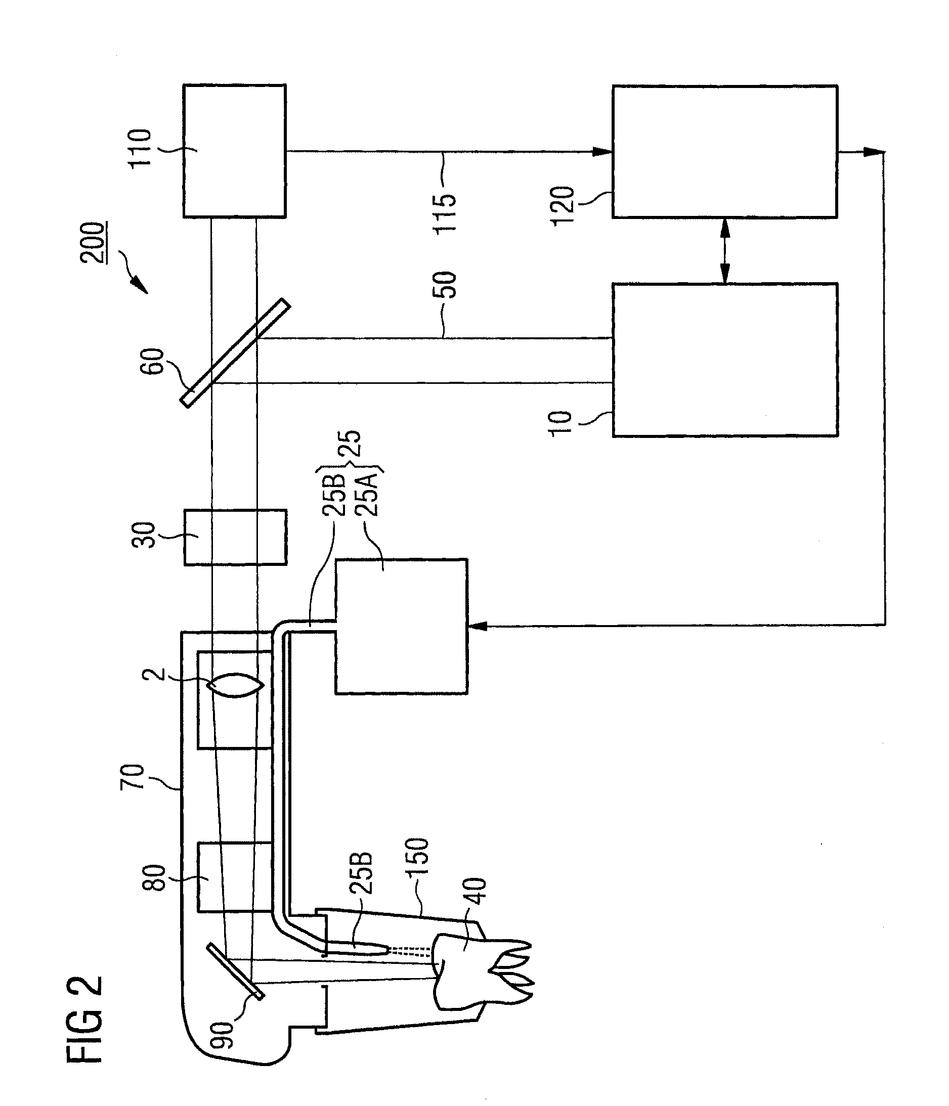
[0035]FIG. 1 illustrates one embodiment of a laser device for lasering biological tissue, but not to true scale. FIG. 1 shows a dental laser device for lasering, abrading or ablating dentin, particularly carious dentin. However, the laser device may be any other kind of medical laser device for lasering some other kind of biological tissue.

[0036]The laser device 100 comprises a laser beam source 1 that may emit a pulsed laser beam 50 with a laser pulse ranging from 1 to 100 ps. The laser beam may be focused on a patient's tooth 4. It may be necessary to first deflect the laser beam with an optical diverter 3 such as a mirror or deviation prism.

[0037]The laser beam source 1 may generate the laser pulses so that the energy per pulse does not exceed 100 pJ. In this case, the focuser 2 for maintaining the energy density values is set so that the laser beam is focused on the surface of the tooth 4 with a diameter range from 10 to 100 μm. The laser beam source 1 may emit the laser pulses ...


PUM


Abstract

Device and method for lasering biological tissue. In a general aspect, the device for lasering a biological tissue may include a source configured to provide a pulsed laser beam, an outcoupler configured to couple the laser beam towards the tissue, and an outfeeder configured to feed a photosensitizer in a direction of the tissue where the outfeeder is connected to the outcoupler. In another general aspect, a method for lasering a biological tissue may include applying a photosensitizer towards the tissue, providing a pulsed laser beam, and lasering a site of the tissue with the pulsed laser beam where the laser beam is emitted with a temporal width at a half maximum range from about 1 ps to about 100 ps.

Description

CROSS-REFERENCE TO RELATED APPLICATIONS[0001]This application is a U.S. continuation application filed under 37 C.F.R. 1.53(b) claiming priority benefit of U.S. application Ser. No. 13 / 064,313, filed Mar. 17, 2011, pending. This application is a continuation application and claims the benefit under 35 U.S.C. Section 111(a), of PCT International Application No. PCT / EP2009 / 006021, filed Aug. 19, 2009, which claimed priority to German Application No. DE 10 2008 047 640.4, filed Sep. 17, 2008, the disclosures of which are incorporated herein in its entirety.BACKGROUND[0002]1. Field[0003]The invention relates to a device and method for lasering biological tissue, and more particularly, to devices and methods for instant diagnosis and lasering biological tissue.[0004]2. Description of the Related Art[0005]One example of the field of application of the invention is dentistry. In dentistry, a method and a corresponding laser device can be used instead of a mechanical drill for the ablation ...

Claims


Application Information

Patent Timeline
Patent Type & Authority Applications(United States)
IPC IPC(8): A61C17/00A61B18/20
CPCA61B18/20A61B2018/2035A61N5/062A61C1/0046A61B2018/2085A61B2018/20351A61C1/00A61C3/02
Inventor KASENBACHER, ANTON
Owner LUMERA LASER
Why Eureka
Try Eureka