Method and system for detecting cardiac arrhythmia

a cardiac arrhythmia and detection method technology, applied in the field of physiological monitoring and diagnosis, can solve the problems of erroneous back-up beat, high chaotic sequence of heartbeats, and typically too small repolarization signal to be seen

Inactive Publication Date: 2012-08-16
WIDEMED
View PDF10 Cites 24 Cited by
  • Summary
  • Abstract
  • Description
  • Claims
  • Application Information

AI Technical Summary

Problems solved by technology
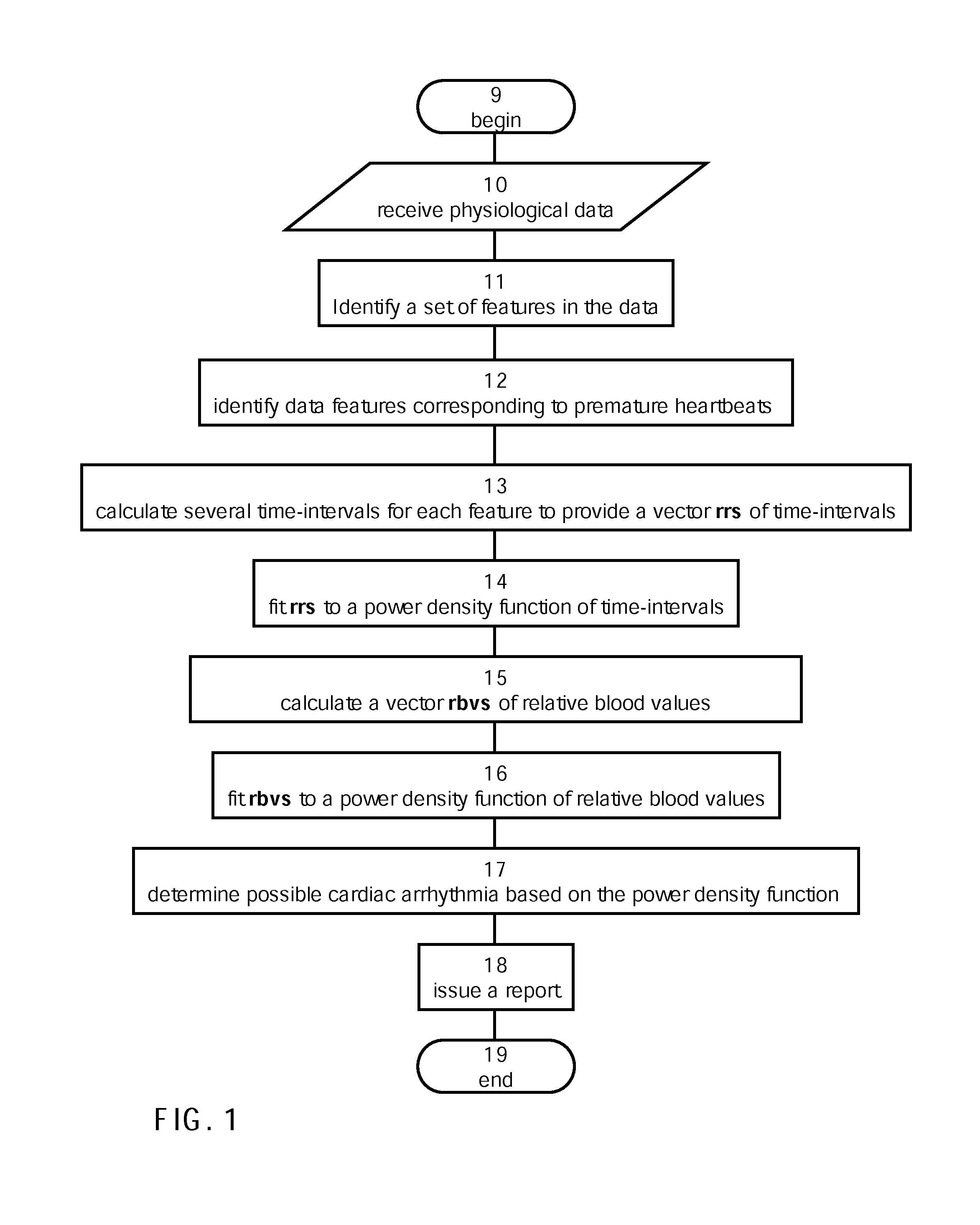
However, from a surface ECG, this repolarization signal is typically too small to be seen.
While one would ideally measure the “ventricular rate” as the QQ interval (typically the interval from QRS onset to the next QRS onset), in practice, the RR interval is used as the measurement of ventricular rate, due to the practical difficulty of reliably measuring the small, inconsistently sized and inconsistently occurring Q-wave.
However, premature contractions occur before the sinus node and override the correct sinus beat, thus indicating a problem with the area of the heart that prematurely generated an erroneous back-up beat.
When ectopics become frequent, it is usually caused by a specific part of the heart causing a problem.
Because the area of the heart that generates the next heartbeat is not fixed, the heart rate of the next heartbeat is also not fixed and thus a highly chaotic sequence of heartbeats is observed.

Method used


Image

  • Method and system for detecting cardiac arrhythmia
  • Method and system for detecting cardiac arrhythmia
  • Method and system for detecting cardiac arrhythmia

Examples


example 1

[0131]Embodiments of the present embodiments have been utilized to detect arrhythmias from ECG and PPG data

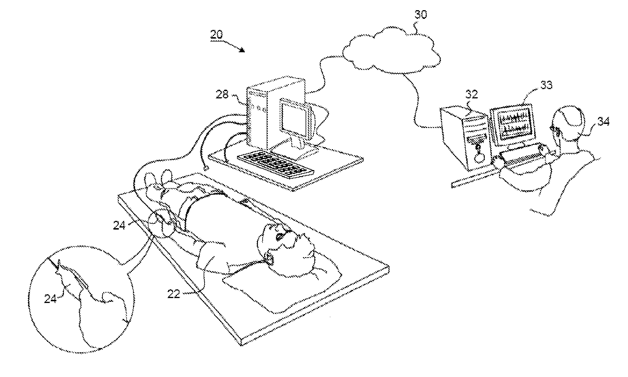
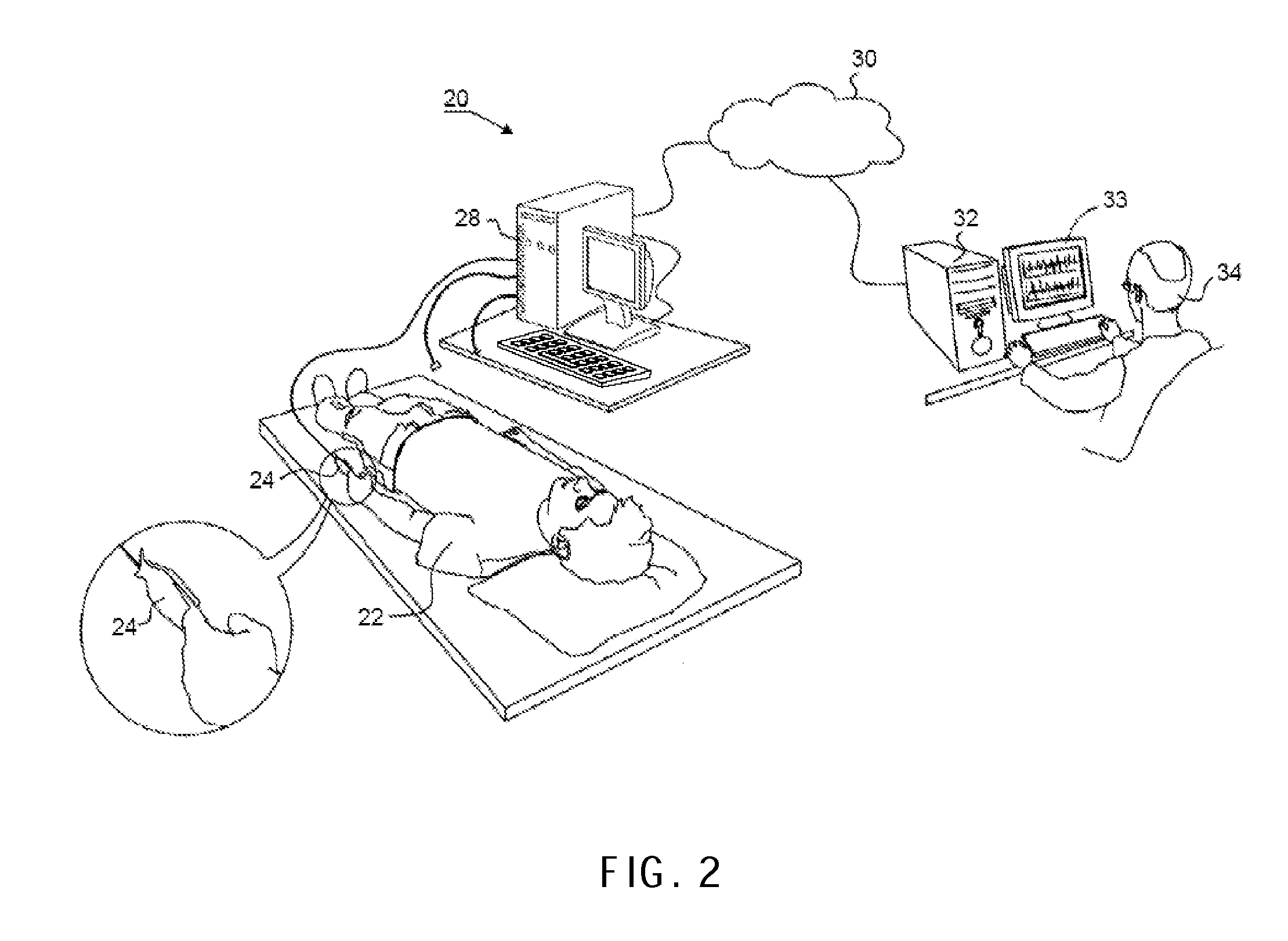
[0132]64 eligible patients were recruited from the Heart Failure Center at Lady Davis Carmel Medical Center / Lin Medical Center in Haifa, Israel. Patients were followed and treated for symptomatic HF of at least 1 month's duration. Treatment was according to AHA / ACC guidelines. Only patients willing to sign an informed consent were enrolled in the study. The study was approved by the medical center's Helsinki Committee. Included were 64 HF patients (54 men and 10 women; age range 27-88 years). The patient underwent full-night PSG sleep test, which include 12 leads ECG and PPG channel on top of the standard channels of PSG test i.e. EEG, chin EMG, EOG, LEG, oximetry, respiratory effort and respiratory flow.

[0133]Additional ECG data were obtained from Moody et al. supra and Goldberger et al. “PhysioBank, PhysioToolkit and PhsioNet: Components of a new Research Resource for Complex...

example 2

[0149]Embodiments of the present embodiments have been utilized to detect premature heartbeats. ECG and PPG data were obtained, preprocessed and the ventricular depolarization were identified as described in Example 1 above.

[0150]The series RT[n] and RV[n] were calculated from rr1[n] and rbv1[n] as described above.

[0151]FIGS. 8A-B shows a representative example of a premature ventricular contraction beat as it is manifested on the ECG (FIG. 8A) and the PPG (FIG. 8B). As shown in both figures, the QRS of the premature ventricular contraction is wide and has no existing P wave. Additionally, the premature ventricular contraction has prolonged compensatory pause before the next heartbeat appears. As shown in FIG. 8B, the relative blood volume value of the premature ventricular contraction is 0.1, the relative blood volume of a normal beat (e.g., a beat preceding the premature ventricular contraction) is 1, and the relative blood volume of the beat immediately following the compensatory...


PUM


Abstract

A method of analyzing physiological data indicative of myocardial activity is disclosed. The method comprises: identifying in the data a set of N features, each corresponding to a ventricular depolarization, and calculating M time-intervals for each ventricular depolarization feature, thereby providing a vector of N*M time-intervals. The method further comprises fitting the vector to a power density function of time-intervals, and determining possible cardiac arrhythmia based on statistical parameters characterizing the function.

Description

RELATED APPLICATIONS[0001]This application claims the benefit of priority from U.S. Patent Application Nos. 61 / 253,110 filed Oct. 20, 2009, and 61 / 254,704 filed Oct. 25, 2009, the contents of which are hereby incorporated by reference as if fully set forth herein.FIELD AND BACKGROUND OF THE INVENTION[0002]The present invention, in some embodiments thereof, relates to physiological monitoring and diagnosis and, more particularly, but not exclusively, to a method and system for detecting cardiac arrhythmia. Some embodiments of the present invention relate to classification of heart beats.[0003]Cardiac arrhythmias can generally divide into life-threatening arrhythmias such as ventricular fibrillation and tachycardia, and non-life-threatening arrhythmias that are not imminently life threatening, such as atrial fibrillation, atrial flutter, ventricular bigeminy and ventricular trigeminy. By way of background, non-life-threatening arrhythmias will now be explained.[0004]In cardiac practic...

Claims


Application Information

Patent Timeline
Patent Type & Authority Applications(United States)
IPC IPC(8): A61B5/0452A61B6/00A61B5/021A61B5/02A61B5/332A61B5/352
CPCA61B5/0059A61B5/02416A61B5/726A61B5/0404A61B5/0456A61B5/04011A61B5/341A61B5/332A61B5/352
Inventor AMOS, YARIV AVRAHAMKAMINSKI, GIL
Owner WIDEMED
Why Eureka
Try Eureka