Productions of artificial tissues by means of tissue engineering using agarose-fibrin biomaterials
a technology of agarose-fibrin and tissue engineering, which is applied in the direction of prosthesis, drug composition, nervous system cells, etc., can solve the problems of limited artificial bladder models available for treating patients needing them, the problem of increasing the risk of recurrence, and the inability to meet the threshold stress, etc., to achieve the effect of improving the threshold stress (in pascal)
- Summary
- Abstract
- Description
- Claims
- Application Information
AI Technical Summary
Problems solved by technology
Method used
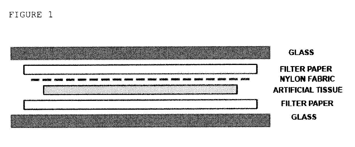
Image

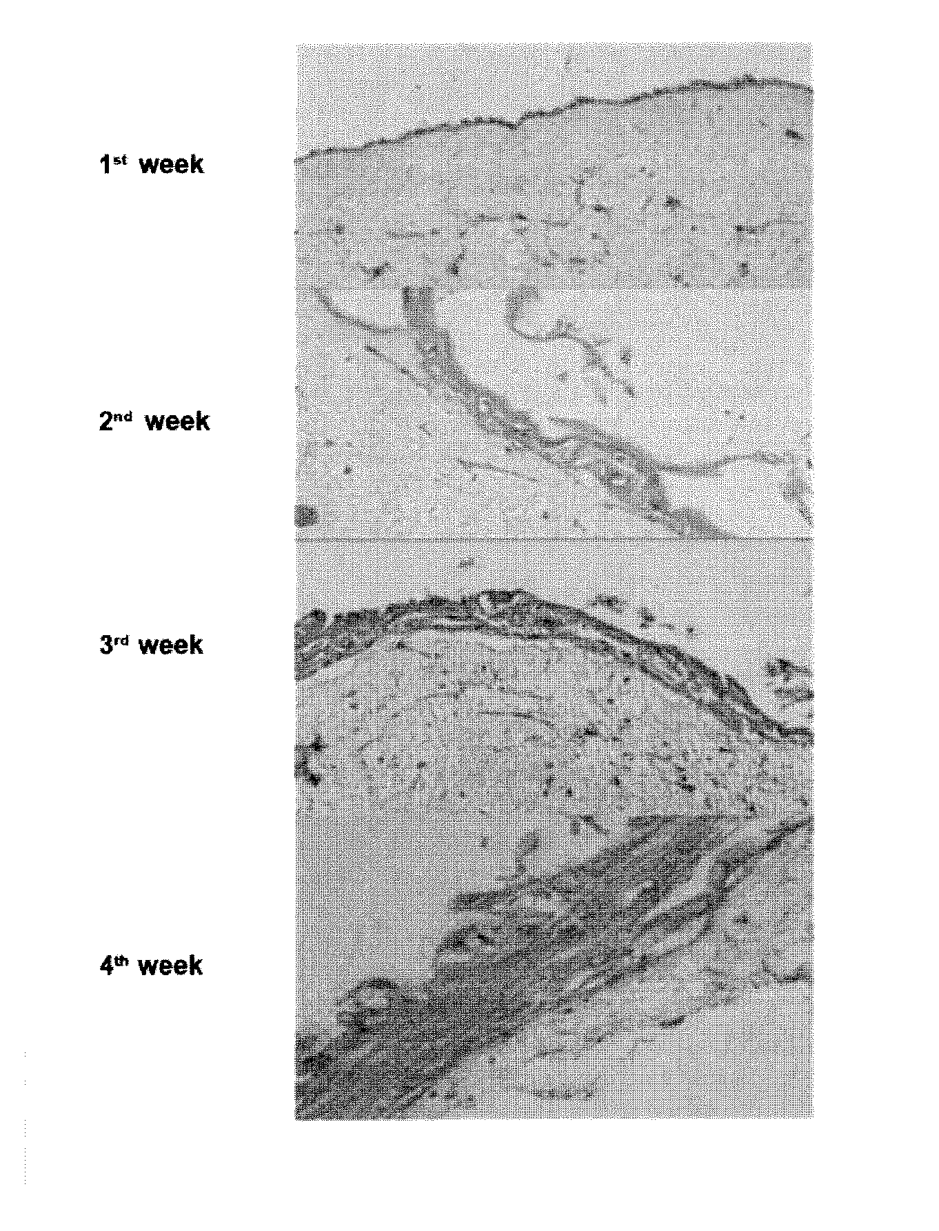
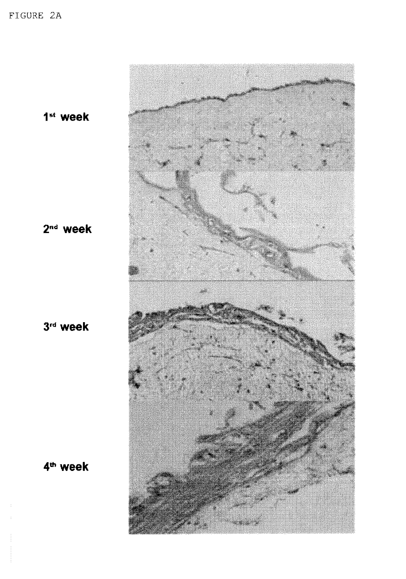
Examples
example 1
Protocol for Preparing an Artificial Human Skin Product
A.—Obtaining Human Skin Samples.
[0170]Full thickness skin samples obtained from donors under local and locoregional anesthesia are used. Once the sample is sterilely obtained, the subcutaneous fatty tissue will be removed wills the aid of scissors until exposing the dermis layer. The removed tissues will then be immediately introduced in a sterile transport medium made up of Dulbecco's Modified Eagle Medium (DMEM) supplemented with antibiotics (500 U / ml of penicillin G and 500 μg / ml of streptomycin) and antimycotic agents (1.25 μg / ml of amphotericin B) to prevent a possible sample contamination.
B.—Generating Primary Fibroblast and Keratinocyte Cultures.
[0171]After the transport period, all the samples must he washed two times in a sterile PBS solution with penicillin, streptomycin and amphotericin B (500 U / ml, 500 μg / ml and 1.25 μg / ml, respectively) to remove all the blood, fibrin, fat or foreign material residues which may adhe...
example 2
Preparing an Artificial Human Skin Product Using Wharton'S Jelly Stem Cells
A.—Obtaining Skin and Human Umbilical Cord Samples.
[0204]Full thickness skin samples obtained from donors under local and locoregional anesthesia are used. Once the sample is sterilely obtained, the subcutaneous fatty tissue will be removed with the aid of scissors until exposing the dermis layer. The removed tissues will then be immediately introduced in a sterile transport medium made up of Dulbecco's Modified Eagle Medium (DMEM) supplemented with antibiotics (500 U / ml of penicillin G and 500 μg / ml of streptomycin) and antimycotic agents (1.25 μg / ml of amphotericin B) to prevent a possible sample contamination.
[0205]The umbilical cords used are obtained from the cesarean birth of normal term pregnancies. After each birth a 10-15 cm fragment of the umbilical cord is obtained which is immediately taken to the laboratory in a transport medium similar to that used for the skin.
B.—Generating Primary Fibroblast a...
example 4
Preparing an Artificial Human Urethra Product
Protocol for Preparing an Artificial Human Urethra Product:
[0268]A.—Obtaining human urethra samples.
[0269]To generate artificial urethras, small biopsies of normal human urethra obtained by means of endoscopy of normal patients or donors are used. Once the sample is sterilely obtained, the removed tissues will be immediately introduced in a sterile transport medium made up of Dulbecco's Modified Eagle Medium (DMEM) supplemented with antibiotics (500 U / ml of penicillin G and 500 μg / ml of streptomycin) and antimycotic agents (1.25 μg / ml of amphotericin B) to prevent a possible sample contamination.
[0270]Alternatively, in the cases in which taking human urethra samples is not feasible, oral mucosa samples or skin samples can be used to generate urethra replacements.
B.—Generating primary stromal and epithelial cell cultures.
[0271]After the transport period all the samples must be washed two times in a sterile PBS solution with penicillin, str...
PUM
| Property | Measurement | Unit |
|---|---|---|
| concentration | aaaaa | aaaaa |
| concentration | aaaaa | aaaaa |
| concentration | aaaaa | aaaaa |
Abstract
Description
Claims
Application Information
 - R&D Engineer
- R&D Manager
- IP Professional
- Industry Leading Data Capabilities
- Powerful AI technology
- Patent DNA Extraction
Browse by: Latest US Patents, China's latest patents, Technical Efficacy Thesaurus, Application Domain, Technology Topic.
© 2024 PatSnap. All rights reserved.Legal|Privacy policy|Modern Slavery Act Transparency Statement|Sitemap