
Implant delivery instrument

Active Publication Date: 2010-10-26
DEPUY SYNTHES PROD INC
View PDF77 Cites 86 Cited by
  • Summary
  • Abstract
  • Description
  • Claims
  • Application Information

AI Technical Summary

Benefits of technology

[0014]Orthopaedic implants useful in approximating, repair or regeneration of fibrocartilage are disclosed in the following applications for U.S. patent Ser. No. 10/195,794 entitled “Meniscus Regeneration Device and Method”; Ser. No. 10/195,719 entitled “Devices from Naturally Occurring Biologically Derived Materials”; Ser. No. 10/195,347 (now U.S. Pat. No. 7,361,195 issued Apr. 22, 2008), entitled “Cartilage Repair Apparatus and Method”; Ser. No. 10/195,344 (now U.S. Pat. No. 7,163,563 issued Jan. 16, 2007), entitled “Unitary Surgical Device and Method”; Ser. No. 10/195,341 entitled “Hybrid Biologic/Synthetic Porous Extracellular Matrix Scaffolds”; Ser. No. 10/195,606 entitled “Cartilage Repair and Regeneration Device and Method”; Ser. No. 10/195,354 entitled “Porous Extracellular Matrix Scaffold and Method”; Ser. No. 10/195,334 entitled “Cartilage Repair and Regeneration Scaffolds and Method”; Ser. No. 10/195,633 (now U.S. Pat. No. 7,201,917 issue

Problems solved by technology

When the soft tissue of a joint is no longer healthy, there can be debilitating pain in the joint.
Soft tissue health can be adversely affected by disease, aging, or trauma.
One form of intra-articular cartilage that is frequently damaged or degenerated is the meniscus of the knee.
The meniscus is frequently damaged in twisting injuries.
It is also damaged with repetitive impact over time.
Meniscus degeneration can also occur by aging; as a person ages, the meniscus can become soft in places, so that even common motions like squatting can cause meniscal tears.
Although menisectomies provide immediate relief to the patient, in the long term the absence of part of the meniscus can cause cartilage wear on the condylar surface, eventually leading to arthritic conditions in the joint.

Method used


Image

  • Implant delivery instrument
  • Implant delivery instrument
  • Implant delivery instrument

Examples


sixth embodiment

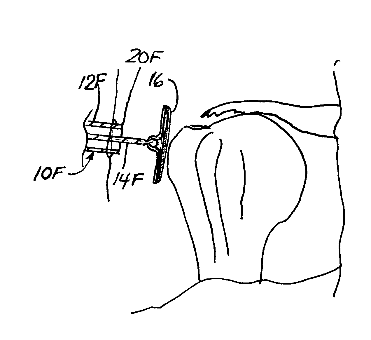
[0101]the surgical instrument of the present invention is illustrated in FIGS. 16-23. In the sixth illustrated instrument 10F, the elongate guide member 12F includes a hollow tube portion 60F and a handle portion 62F at the proximal end 18F of the elongate guide member 12F. The handle portion 62F has an enlarged diameter for the surgeon to hold, while the hollow tube portion 60F has an outer diameter that allows for use in arthroscopic surgical procedures, mini-arthrotomies or open arthrotomies. As best seen in FIGS. 16-17, the hollow tube portion 60F has a J-shaped slot 64F extending from the exterior to the interior surface. The J-shaped slot 64F has two axially-oriented segments 66F, 68F joined at their distal ends by a transverse segment 70F. The J-shaped slot 64F receives a pin 72F fixed to the reciprocable member 14F of the sixth illustrated instrument 10F.

[0102]The reciprocable member 14F of the sixth illustrated instrument 10F comprises an assembly of four components. As bes...

eighth embodiment

[0123]a surgical instrument in accordance with the present invention is illustrated in FIGS. 26-30 at 10H. The eighth illustrated surgical instrument shares many common features with the sixth and seventh illustrated instruments 10F, 10G as described above. The implant carrier 24H of the eighth illustrated instrument 10H has arms 100H with end clips 112H like those of the seventh instrument 10G. The implant carrier portion 24H and the distal rod 94H of the connector member 78H of the eighth illustrated surgical instrument 10H are somewhat different from those of the sixth and seventh illustrated surgical instruments 10F, 10G. In the eighth illustrated surgical instrument 10H, the distal end of the distal rod 94H has flat parallel surfaces 120H. The base 98H of the implant carrier 24H is integral with the arms 100H, and comprises a pair of spaced parallel tabs 122H (see FIGS. 27 and 30) that are placed against the flat parallel surfaces 120H (see FIGS. 27 and 30) of the distal rod 94...


PUM


Abstract

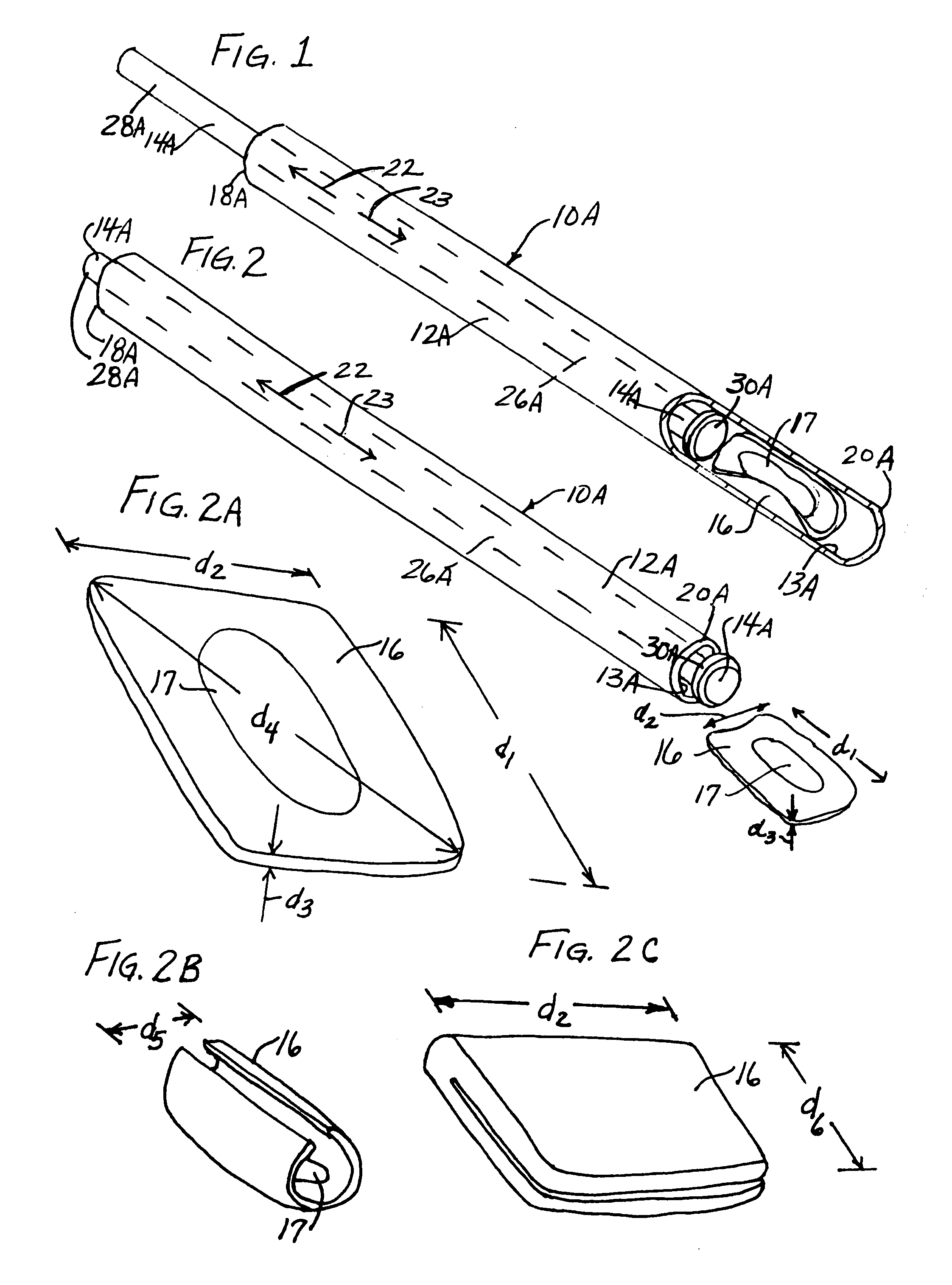
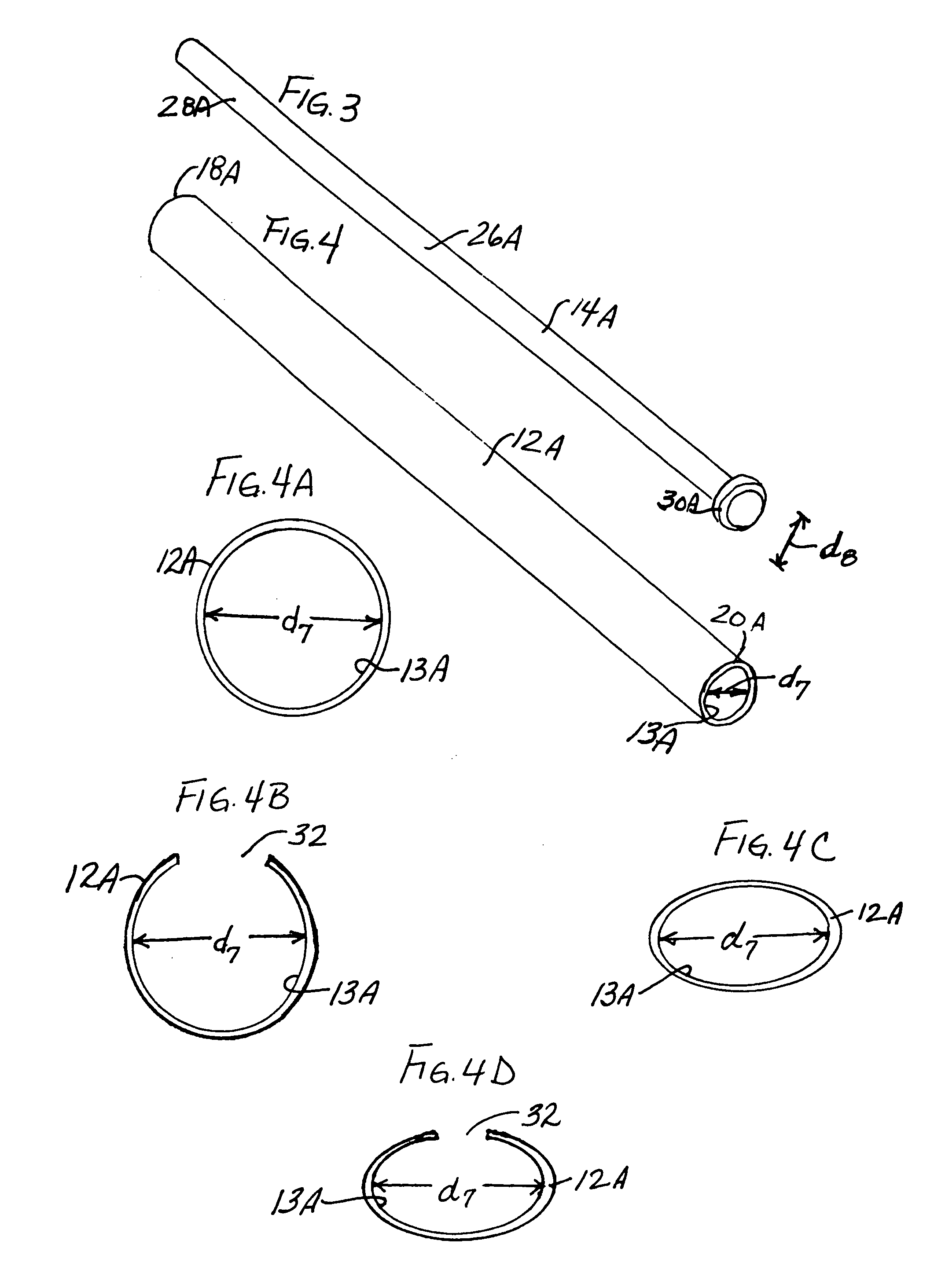
A surgical instrument has an elongate guide member and a reciprocable member. The elongate guide member provides a protected path of travel for an implant from outside the body to a position near a damaged tissue site. The reciprocable member is movable along the elongate guide member to push the implant along its path of travel. The reciprocable member can include an implant carrier. The implant can be an orthopaedic implant for repair or regeneration of soft tissue at a damaged joint site. The implant can be larger than the transverse dimension of the elongate guide member and folded or rolled to fit within the elongate guide member. Use of this instrument protects the implant from damage as it is delivered to the damaged joint site. The invention also includes a surgical method for delivering an implant to a damaged tissue site.

Description

FIELD OF THE INVENTION[0001]The present invention relates generally to a surgical instrument for delivering an implant to a damaged tissue site in the human body.BACKGROUND OF THE INVENTION[0002]Several different types of soft tissue are found in human joints. For example, a joint can include articular hyaline cartilage, intra-articular fibrocartilage, tendons and ligaments. Articular hyaline cartilage is found on the surfaces of the bones of the joint. Intra-articular cartilage is found between the joint surfaces. Tendons connect muscle to the bones of the joint, and ligaments connect articular extremities of the bones of the joint.[0003]When the soft tissue of a joint is no longer healthy, there can be debilitating pain in the joint. Soft tissue health can be adversely affected by disease, aging, or trauma. The adverse effects of disease, aging and trauma can be, for example, in the form of a tear in the soft tissue, or in the form of a breakdown, thinning or delamination of the t...

Claims


Application Information

Patent Timeline
IPC IPC(8): A61F2/00A61B17/16A61B17/88A61F2/30A61F2/38A61F2/46
CPCA61F2/4618A61B17/1635A61B17/8872A61F2/3872A61F2002/30153A61F2002/30235A61F2002/30373A61F2002/30565A61F2002/30579A61F2002/30581A61F2002/30601A61F2002/4635A61F2220/0033A61F2230/0019A61F2230/0069A61F2002/4627A61F2002/4628Y10S606/914
Inventor ZANNIS, ANTHONY D.CAMINO, THOMAS S.KEMPPAINEN, JOHN W.SCHWARTZ, HERBERT E.MCADAMS, DANNY E.
Owner DEPUY SYNTHES PROD INC
Learn More