Methods and Crosslinked Polymer Compositions for Cartilage Repair

a crosslinked polymer and cartilage technology, applied in the field of cartilage repair methods, can solve the problems of not being readily degraded in vivo, and achieve the effect of facilitating tissue healing and regeneration

Inactive Publication Date: 2009-05-07
ANGIOTECH PHARMA US
View PDF4 Cites 67 Cited by
  • Summary
  • Abstract
  • Description
  • Claims
  • Application Information

AI Technical Summary

Benefits of technology

[0009]In one aspect of the invention, the compositions can be used for repairing injured cartilage tissue (e.g., articular or meniscal cartilage) at a treatment site. The treatment site may be, for example, in a joint (e.g., in the knee, shoulder, ankle, elbow, wrist, and the like) and the compositions may be used for the repair of articular cartilage defects and / or meniscal tears. The described compositions may facilitate growth of new cartilage tissue at the site of implantation. The described compositions can be used to facilitate attachment of connective tissue, such as cartilage, to the underlying bone. The described compositions can also facilitate the attachment of other connective tissues such as tendon, ligaments, fat, muscle or other tissue to underlying bone or periosteum during procedures such as facelifts, tendon and ligament repairs, soft tissue reconstructive procedures, and cosmetic implant procedures (such as breast implants and facial implants).
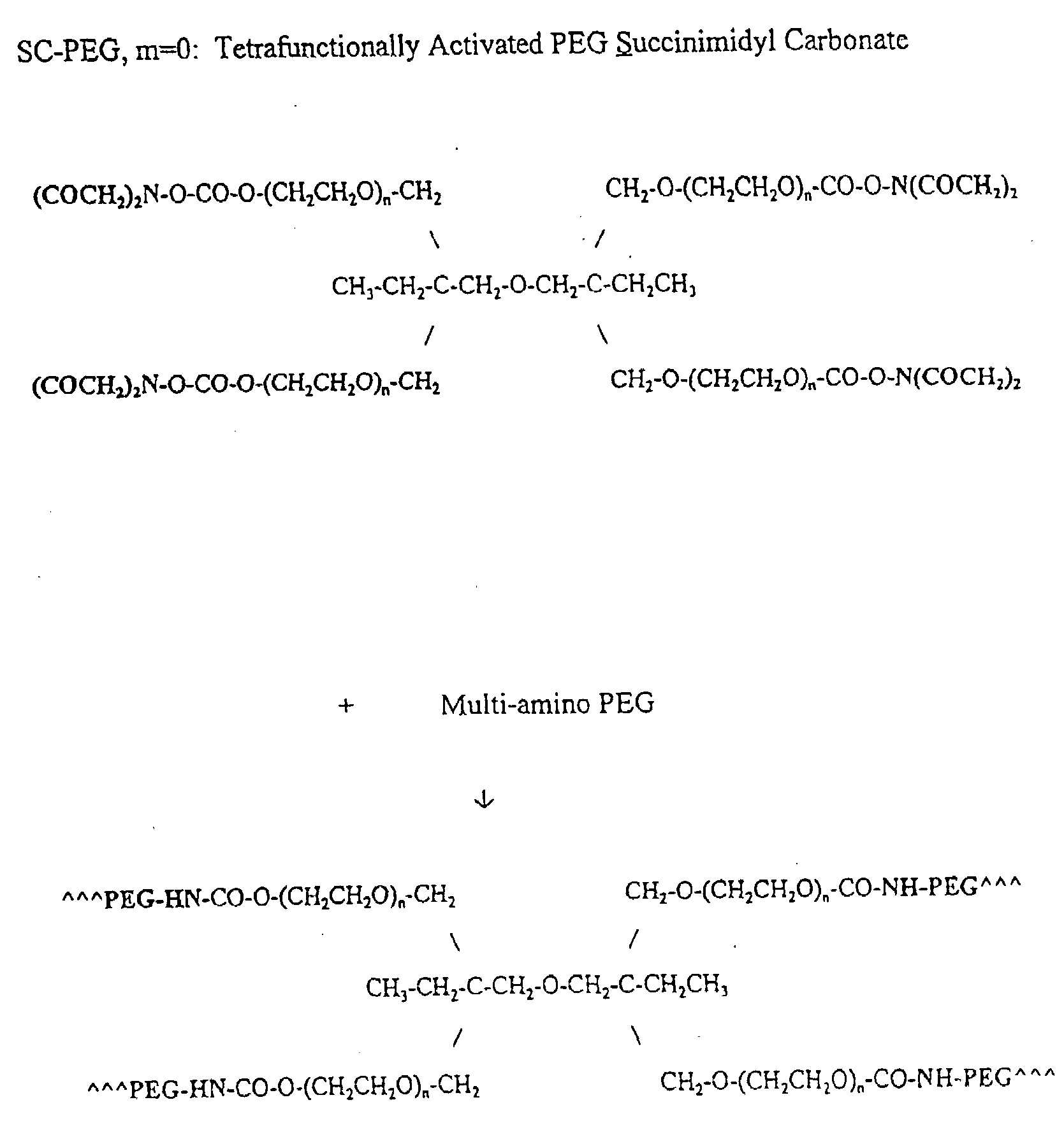
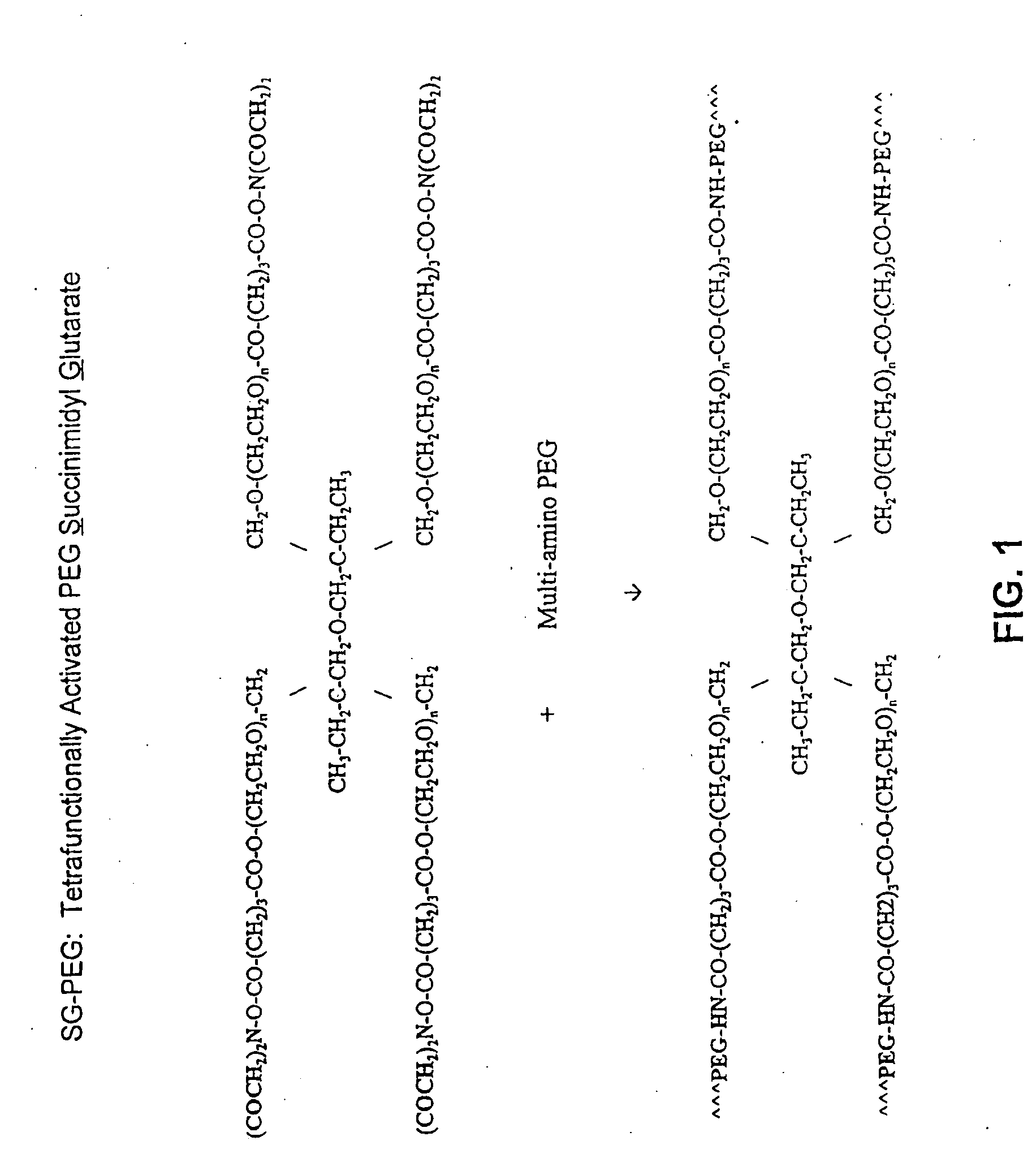
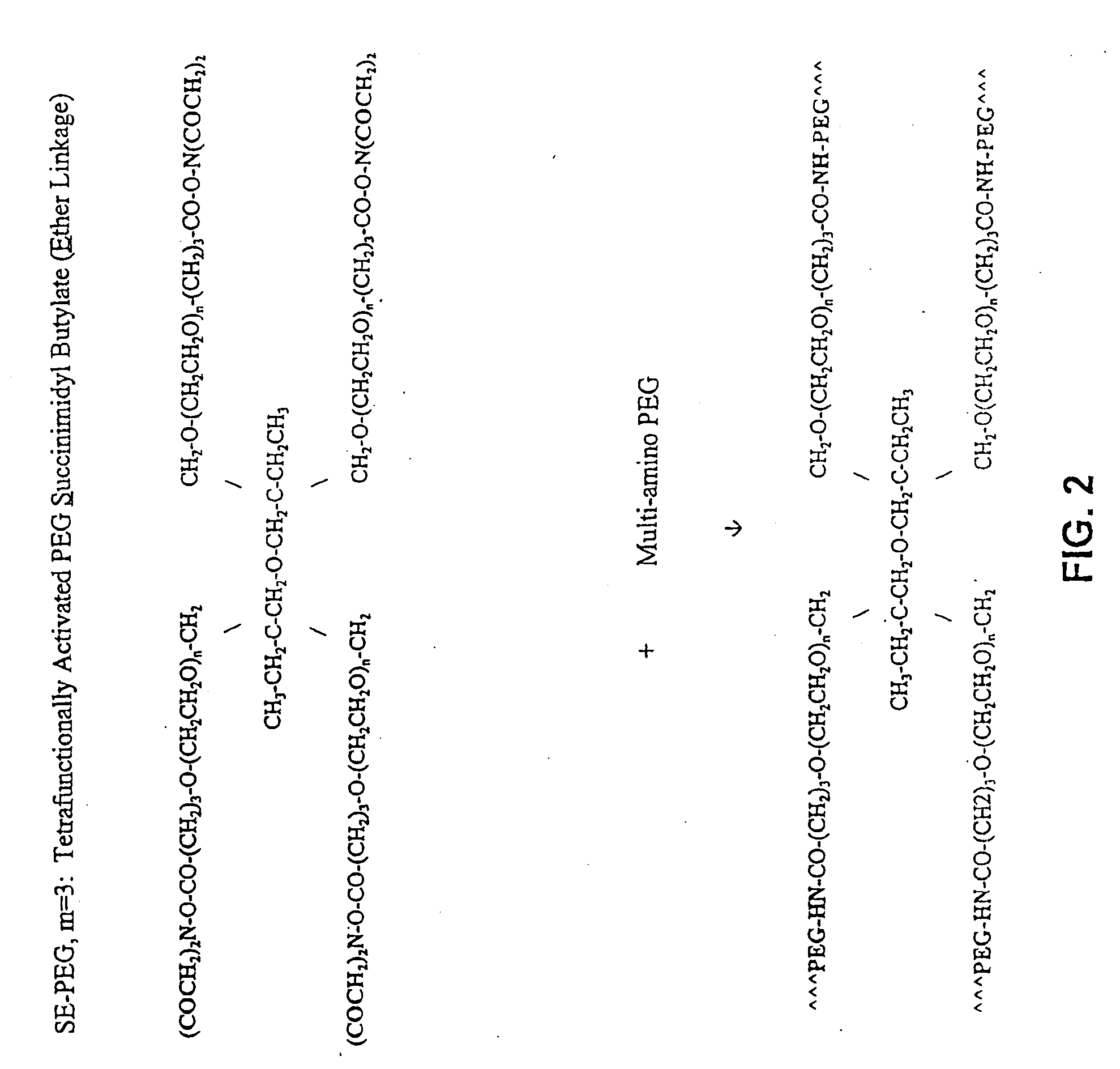
[0011]The composition has a hydrophilic polymer and crosslinkable components that may be readily crosslinked upon admixture with an aqueous medium to provide a crosslinked composition suitable for use as a biomaterial. The composition is biocompatible, and does not leave any toxic, inflammatory, or immunogenic reaction products at the site of administration. Furthermore, as the composition is not subject to enzymatic cleavage by matrix metalloproteinases such as collagenase, it is not readily degradable in vivo. As a result, the composition may degrade more slowly than either the hydrophilic polymer component or the crosslinkable component as the two components will serve to mutually protect each other from the effects of metalloproteases or hydrolysis.
[0046]In the aforementioned methods for repairing damaged cartilage tissue, damaged connective tissue, and / or damaged soft tissue in a patient, the compositions may comprise a biologically active agent, which may be delivered to the damaged cartilage tissue, damaged connective tissue, and / or damaged soft tissue in the patient. For delivery to the tissues, the composition may be in the form of microparticles, nanoparticles, microemulsions, emulsions, liposomes and micelles. A preferred biologically active agent for use in the methods of the present invention is an angiogenesis inhibitor. Another preferred biologically active agent for use in the methods of the present invention is paclitaxel or an analogue or derivative thereof. Additionally, other biologically active agents incorporated in the compositions of the present invention may facilitate tissue healing and regeneration.

Problems solved by technology

Furthermore, as the composition is not subject to enzymatic cleavage by matrix metalloproteinases such as collagenase, it is not readily degradable in vivo.

Method used


Image

  • Methods and Crosslinked Polymer Compositions for Cartilage Repair
  • Methods and Crosslinked Polymer Compositions for Cartilage Repair
  • Methods and Crosslinked Polymer Compositions for Cartilage Repair

Examples


example 1

Gelation OF NHS-PEG and HS-PEG

[0264]A homogenous mixture of NHS-PEG and HS-PEG was obtained by mixing approximately equal amounts of the two powders. A 20% (w / v) solution of mixed PEG powder was then prepared by dissolving the powder in diluted HCl. The obtained solution (pH 2.1) was then cosprayed with an equal volume of a 300 mM sodium phosphate / sodium carbonate buffer (pH 9.6). Gelation occurred almost immediately (<3 sec) and the gel obtained its firm, rubbery, solid properties in less than a minute with a total solid content of approximately 10% PEG.

example 2

Gelation of NHS-PEG, HS-PEG, and Methylated Collagen

[0265]A crosslinked hydrogel was formed from an acidic solution of NHS-PEG, HS-PEG, and a methylated collagen (MC). The acidic paste (pH 3-4) containing 10% NHS-PEG, 10% HS-PEG, and 22 mg / mL of MC spontaneously gelled when mixed with an equal volume of 0.3 M phosphate / carbonate solution (pH 9.6). Gelation occurred within seconds to form a strong hydrogel that adheres well to the tissue and swells in a controlled fashion in saline solution. As previously noted, the combination of NHS-PEG (10K), HS-PEG (10K), and MC is associated with the tradename CHONDROGEL™.

example 3

Gelation of NHS-PEG, HS-PEG, and MC on a Tissue Sample

[0266]A crosslinked hydrogel (i.e., CHONDROGEL™) was formed from an acidic solution of NHS-PEG, HS-PEG, and a methylated collagen (MC). The acidic paste (pH 3-4) containing 10% NHS-PEG, 10% HS-PEG, and 22 mg / mL of MC was applied onto a tissue and shaped with a spatula to its desired form. In a subsequent step, a smaller volume of 0.3 M phosphate / carbonate solution (pH 9.6) than that used in Example 2 was applied (via spraying or dropping) to the paste. Gelation of the paste started immediately and continued as the 0.3 M phosphate / carbonate solution diffused into the tissue. Within a few minutes, a strong hydrogel that adhered well to the tissue and swells in a controlled fashion in saline solution was formed on the tissue.


PUM

PropertyMeasurementUnit
biocompatibleaaaaaaaaaa
hydrophilicaaaaaaaaaa
hydrophobicaaaaaaaaaa

Abstract

A method of repairing damaged cartilage and soft tissue in a patient is provided using a biocompatible, non-immunogenic composition. The composition comprises a hydrophilic polymer and a plurality of crosslinkable components having reactive functional groups. The composition used in the method may be loaded with biologically active agents for delivery to the damaged tissues. Kits for use in carrying out the method of the invention are also provided.

Description

CROSS-REFERENCE TO RELATED APPLICATIONS[0001]This application, filed on Dec. 20, 2006, claims priority under 35 U.S.C. § 119(a) to PCT / US2005 / 022343, filed on Jun. 23, 2005, which claims priority under 35 U.S.C. § 119(e) to 60 / 582,651, filed on Jun. 23, 2004, both of which are incorporated by reference in their entireties herein.TECHNICAL FIELD[0002]This invention relates generally to methods for repairing connective tissue (e.g., cartilage) using compositions comprised of a hydrophilic polymer and crosslinked biomaterials.BACKGROUND OF THE INVENTION[0003]Successful human athletic performance requires the optimal function of our articulations. Proper joint function requires not only adequate strength and stability, but a smooth, gliding articular surface to allow an effortless range of motion. The tissue most responsible for creating the articular surface is hyaline cartilage; a tissue composed of chondrocytes (cartilage cells), extracellular matrix (type II collagen, aggrecan, glyc...

Claims


Application Information

Patent Timeline
Patent Type & Authority Applications(United States)
IPC IPC(8): A61K31/715A61K38/19A61K31/765A61K31/337A61K35/28A61P19/04A61K39/395A61K38/43A61K38/39
CPCA61L2430/06A61L27/26A61P19/02A61P19/04
Inventor DANILOFF, GEORGE Y.TOLEIKIS, PHILIP M.HUNTER, WILLIAM L.AVELAR, RUI
Owner ANGIOTECH PHARMA US
Why Eureka
Try Eureka