Automatic or Semi-Automatic Whole Body MR Scanning System

Inactive Publication Date: 2012-11-08
SIEMENS HEALTHCARE GMBH
View PDF54 Cites 17 Cited by
  • Summary
  • Abstract
  • Description
  • Claims
  • Application Information

AI Technical Summary

Problems solved by technology

However, continuous patient movement tends to generate a large amount of images that need be read and interpreted, typically through a labor intensive process, by a highly trained health care provider.
Although there is dedicated software available to reduce the workload and shorten the reading time, such software is limited to research applications or specific clinical applications.

Method used


Image

  • Automatic or Semi-Automatic Whole Body MR Scanning System
  • Automatic or Semi-Automatic Whole Body MR Scanning System
  • Automatic or Semi-Automatic Whole Body MR Scanning System

Examples


Embodiment Construction

[0015]A comprehensive and fully integrated system according to invention principles integrates use of patient specific diagnostic data into an imaging session and optimizes MR imaging by minimizing risk of missing necessary diagnostic scans or rescheduling of patients for imaging. The system comprehensively combines MR screening, identification of pathology, diagnostic scans and reporting and reduces table and scan time of the patient as well as the risk of missing relevant diagnostic scans. The system also reduces dependency of image quality on operator experience. The system further streamlines reading and interpretation of images by providing guidance and automatically reporting relevant findings.

[0016]The system automatically identifies cancer patients with metastatic tumor growth regions for subsequent surgery or cancer staging and excludes certain pathologies to identify potential causes of patient symptoms. The system triggers relevant diagnostic image acquisition and reporti...


PUM


Abstract

A system includes an image data processor for automatically processing data representing multiple patient anatomical images acquired in a single imaging scan. The images are acquired by, identifying multiple different anatomical elements in corresponding multiple different anatomical regions and identifying multiple different potentially pathology indicative features associated with the multiple different anatomical elements in response to first predetermined information associating different potentially pathology indicative features with corresponding different anatomical elements. The image data processor determines multiple different image acquisition methods for use in imaging the multiple different potentially pathology indicative features in response to second predetermined information associating different image acquisition methods with corresponding identified different pathology indicative features. An output processor collates images for output.

Description

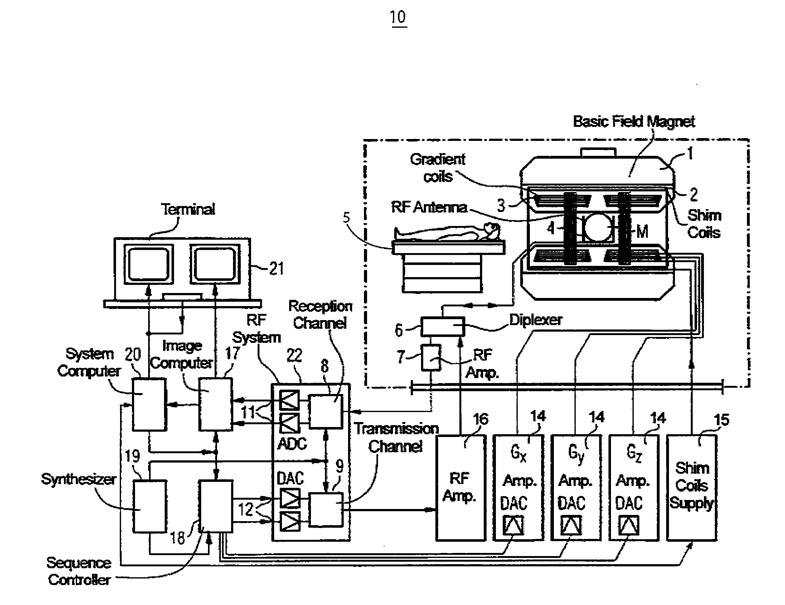
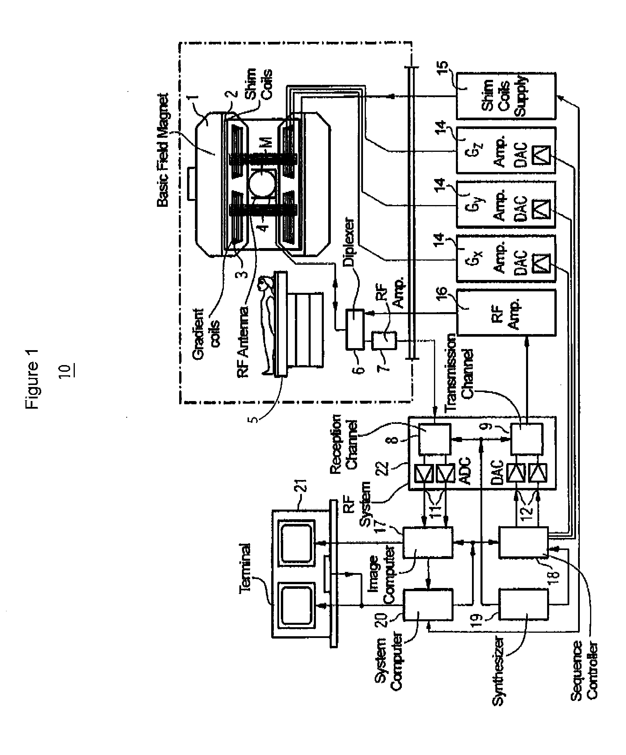
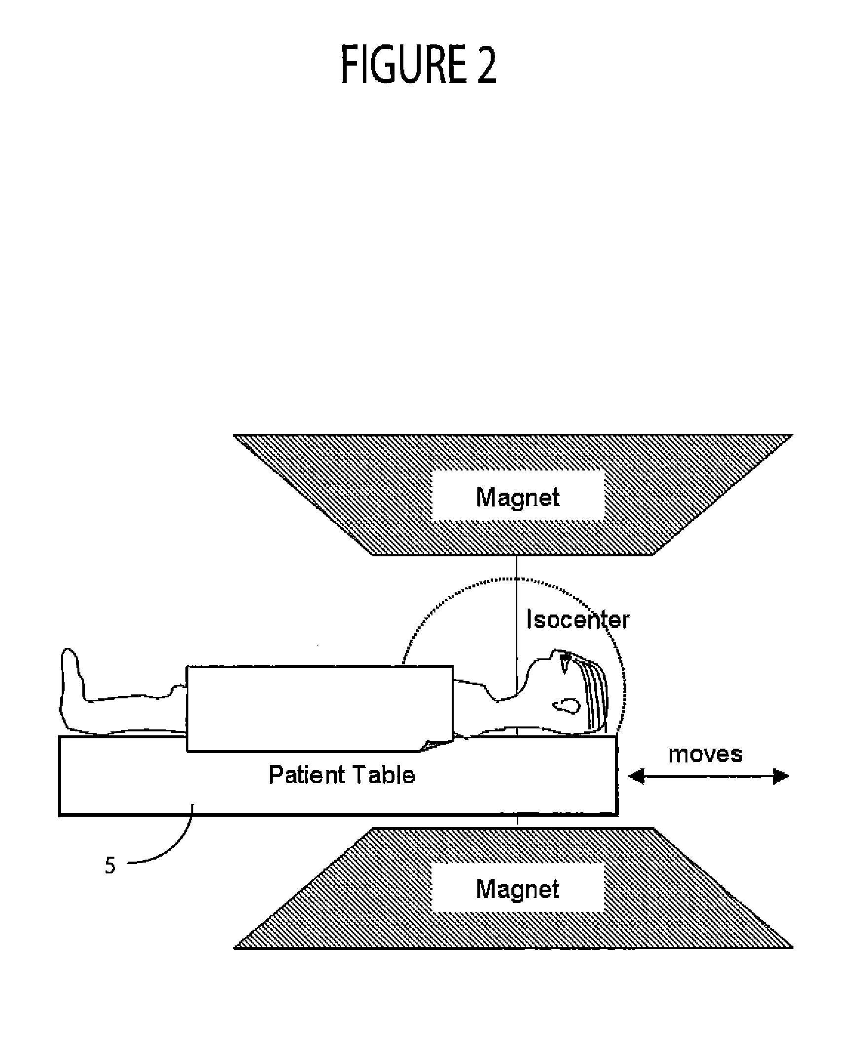
[0001]This is a non-provisional application of provisional application Ser. No. 61 / 482,682 filed 5 May, 2011, by S. Zuehlsdorff et al.FIELD OF THE INVENTION[0002]This invention concerns a system for imaging multiple anatomical regions of a patient by identifying multiple different potentially pathology indicative features and determining multiple different image acquisition methods for use in imaging multiple different potentially pathology indicative features of the regions.BACKGROUND OF THE INVENTION[0003]State of the art whole body MR screening systems often use a continuously moving patient support table during image data acquisition to collect images of an entire patient in an isocenter of an MRI scanner. Typical applications include screening for cancerous metastasis or MR angiographies with and without contrast media. However, continuous patient movement tends to generate a large amount of images that need be read and interpreted, typically through a labor intensive process, ...

Claims


Application Information

Patent Timeline
IPC IPC(8): A61B5/055A61B6/00A61B5/05
CPCA61B5/055A61B6/5217A61B2576/00A61B2505/00A61B2503/12G16H50/30
Inventor ZUEHLSDORFF, SVENGLIELMI, CHRISTOPHERBI, XIAOMING
Owner SIEMENS HEALTHCARE GMBH
Why Eureka
Try Eureka