Method and ultrasound imaging system for image-guided procedures

a technology of ultrasound imaging and image guidance, applied in the field of methods, can solve problems such as difficult and error-prone process of accurately communicating all the relevant information from the endocrinologist to the surgeon

Inactive Publication Date: 2012-11-15
GENERAL ELECTRIC CO
View PDF11 Cites 12 Cited by
  • Summary
  • Abstract
  • Description
  • Claims
  • Application Information

AI Technical Summary

Problems solved by technology

However, since a patient may have multiple lymph nodes that need to be involved in the surgical procedure, accurately co

Method used


Image

  • Method and ultrasound imaging system for image-guided procedures
  • Method and ultrasound imaging system for image-guided procedures
  • Method and ultrasound imaging system for image-guided procedures

Examples


Embodiment Construction

[0013]In the following detailed description, reference is made to the accompanying drawings that form a part hereof, and in which is shown by way of illustration specific embodiments that may be practiced. These embodiments are described in sufficient detail to enable those skilled in the art to practice the embodiments, and it is to be understood that other embodiments may be utilized and that logical, mechanical, electrical and other changes may be made without departing from the scope of the embodiments. The following detailed description is, therefore, not to be taken as limiting the scope of the invention.

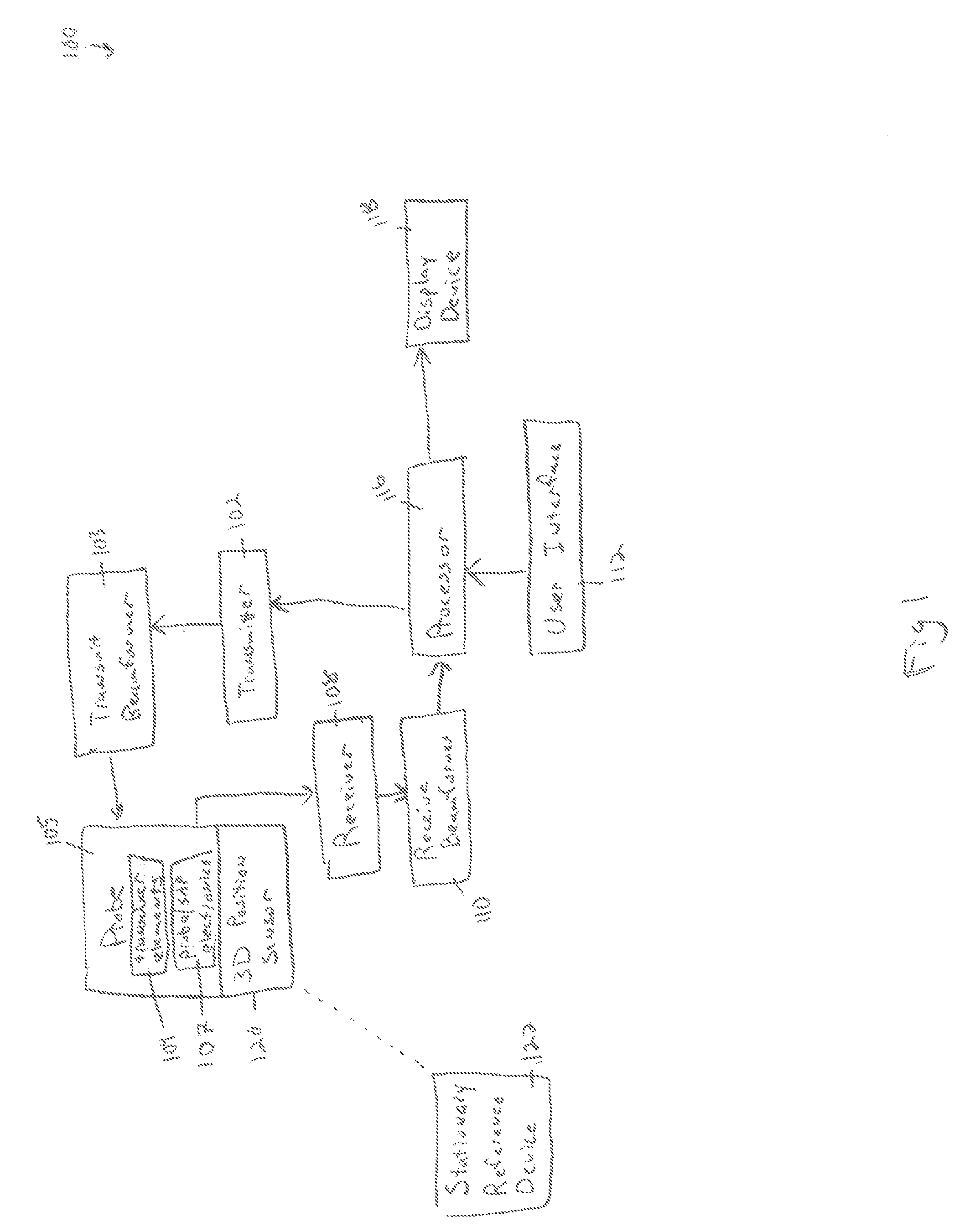
[0014]FIG. 1 is a schematic diagram of an ultrasound imaging system 100 in accordance with an embodiment. The ultrasound imaging system 100 includes a transmitter 102 that transmits a signal to a transmit beamformer 103 which in turn drives transducer elements 104 to emit pulsed ultrasonic signals into a structure, such as a patient (not shown). A probe 105 includes the transd...


PUM


Abstract

A method and ultrasound imaging system for image-guided procedures includes collecting first position data of an anatomical surface with a 3D position sensor. The method and ultrasound imaging system includes generating a 3D graphical model of the anatomical surface based on the first position data. The method and ultrasound imaging system includes acquiring ultrasound data with a probe in position relative to the anatomical surface. The method and ultrasound imaging system includes using the 3D position sensor to collect second position data of the probe in the position relative to the anatomical surface. The method and ultrasound imaging system includes generating an image based on the ultrasound data and identifying a structure in the image. The method and ultrasound imaging system includes registering the location of the structure to the 3D graphical model based on the first position data and the second position data. The method and ultrasound imaging system includes displaying a representation of the 3D graphical model including a graphical indicator of the structure.

Description

FIELD OF THE INVENTION[0001]This disclosure relates generally to a method and ultrasound imaging system for generating a representation of a 3D graphical model for use with image-guided procedures.BACKGROUND OF THE INVENTION[0002]In many areas, it is typical for a diagnostic imaging system operator to acquire images of a planned site for surgery. Then, a surgeon will use the images in order to plan the most appropriate clinical procedure and approach. Using endocrinology as an example, an endocrinologist will usually acquire images of a patient's neck with an ultrasound imaging system in order to identify one or more lymph nodes that are likely to be cancerous. Next, it is necessary for the endocrinologist to communicate the information regarding the precise location of the one or more cancerous lymph nodes to the surgeon. At a minimum, the endocrinologist needs to identify insertion locations for the surgeon. Preferably, the endocrinologist will also communicate information regardi...

Claims


Application Information

Patent Timeline
IPC IPC(8): A61B8/14
CPCA61B8/4254A61B8/4263A61B8/463A61B8/5292A61B8/469A61B8/483A61B8/466A61B8/4455
Inventor HALMANN, MENACHEMWASHBURN, MICHAEL J.
Owner GENERAL ELECTRIC CO
Why Eureka
Try Eureka