Method and apparatus for measuring activity of a tracer

a tracer and activity technology, applied in the field of methods and apparatus for measuring activity of tracers, can solve the problems of inability to measure an image-derived bif from a fixed region, inability to achieve the effect of a fixed region,

Active Publication Date: 2012-12-06
SIEMENS MEDICAL SOLUTIONS USA INC
View PDF3 Cites 14 Cited by
  • Summary
  • Abstract
  • Description
  • Claims
  • Application Information

AI Technical Summary

Benefits of technology

[0014]Thus, instead of the single VOI in a conventional dynamic scan, for a multiple bed position scan the activity values from regions of interest in consecutive data sets can be combined, by modifying the activity values according to the size of a feature in the ROI.

Problems solved by technology

As such, measurement of an image-derived BIF from a fixed region is problematic, if not impossible.
However, the invasiveness of this procedure makes it undesirable for routine use.A reference region-based kinetic model could be used, which does not require a BIF, but instead a VOI placed in a region where no specific binding / uptake of the radiotracer occurs.
Unfortunately, this method is impractical for common tracers such as 18F-FDG where perfused regions lacking specific binding / uptake cannot be identified.

Method used


Image

  • Method and apparatus for measuring activity of a tracer
  • Method and apparatus for measuring activity of a tracer
  • Method and apparatus for measuring activity of a tracer

Examples


Embodiment Construction

[0031]When the following terms are used herein, they have the following definitions.

[0032]PET Positron Emission Tomography

[0033]ROI / VOI Region / volume of Interest

[0034]SUV Standard Uptake Value

[0035]CT Computed Tomography

[0036]BIF Blood Input Function

[0037]FOV Field of View

[0038]PSF Point Spread Function

[0039]PVE Partial Volume Effect

[0040]TAC Time Activity Curve

[0041]WB Whole Body

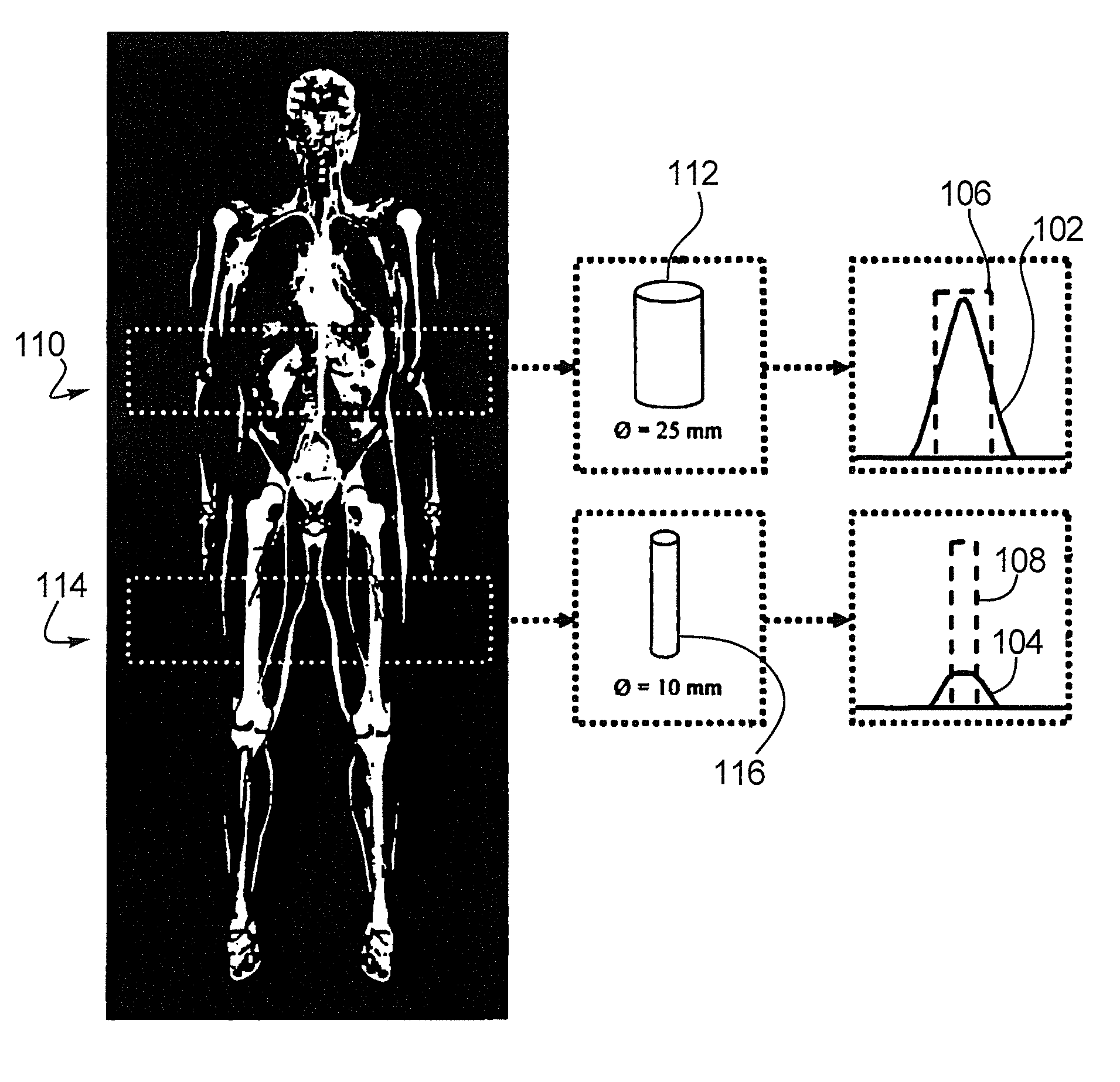
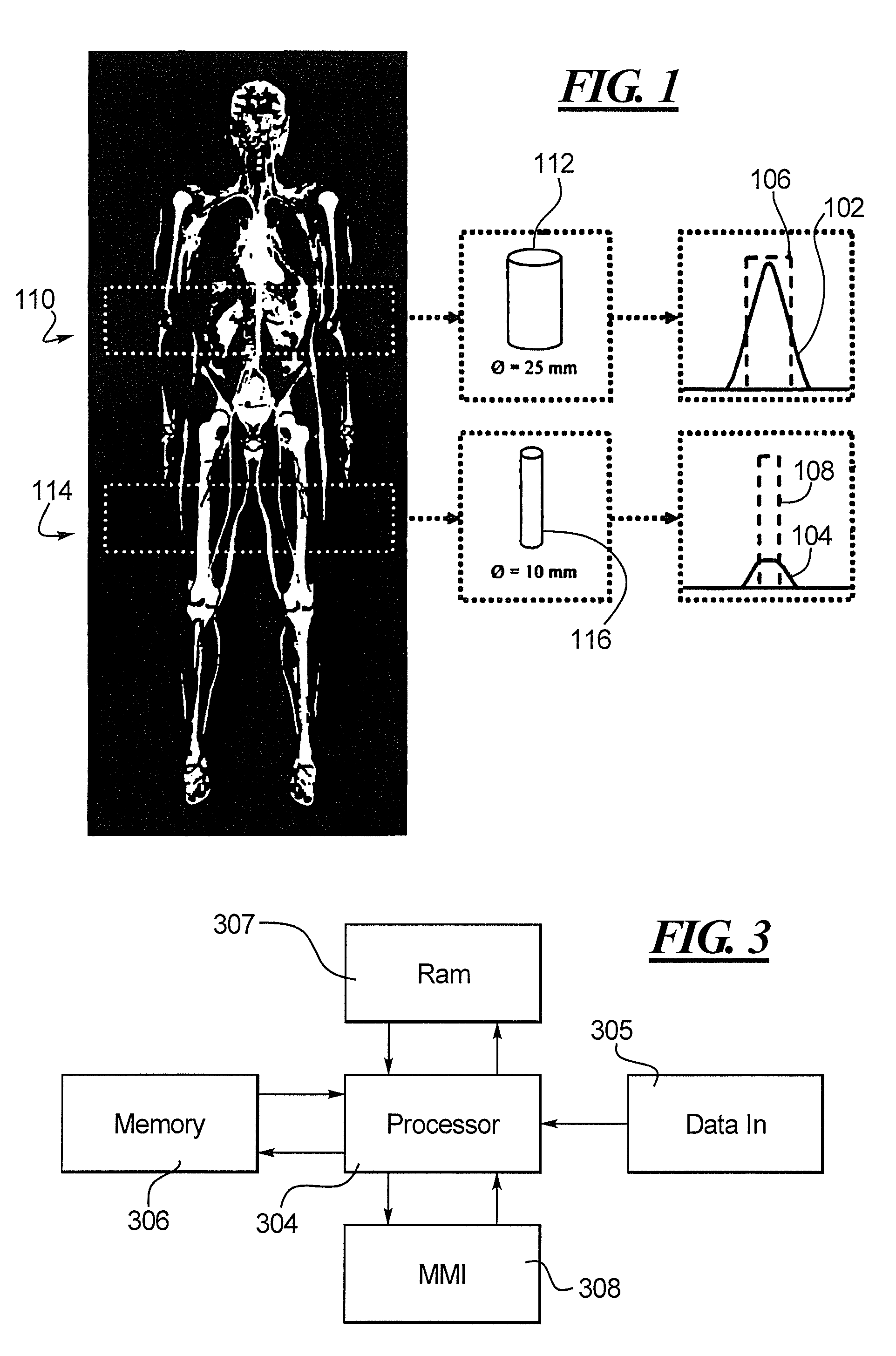
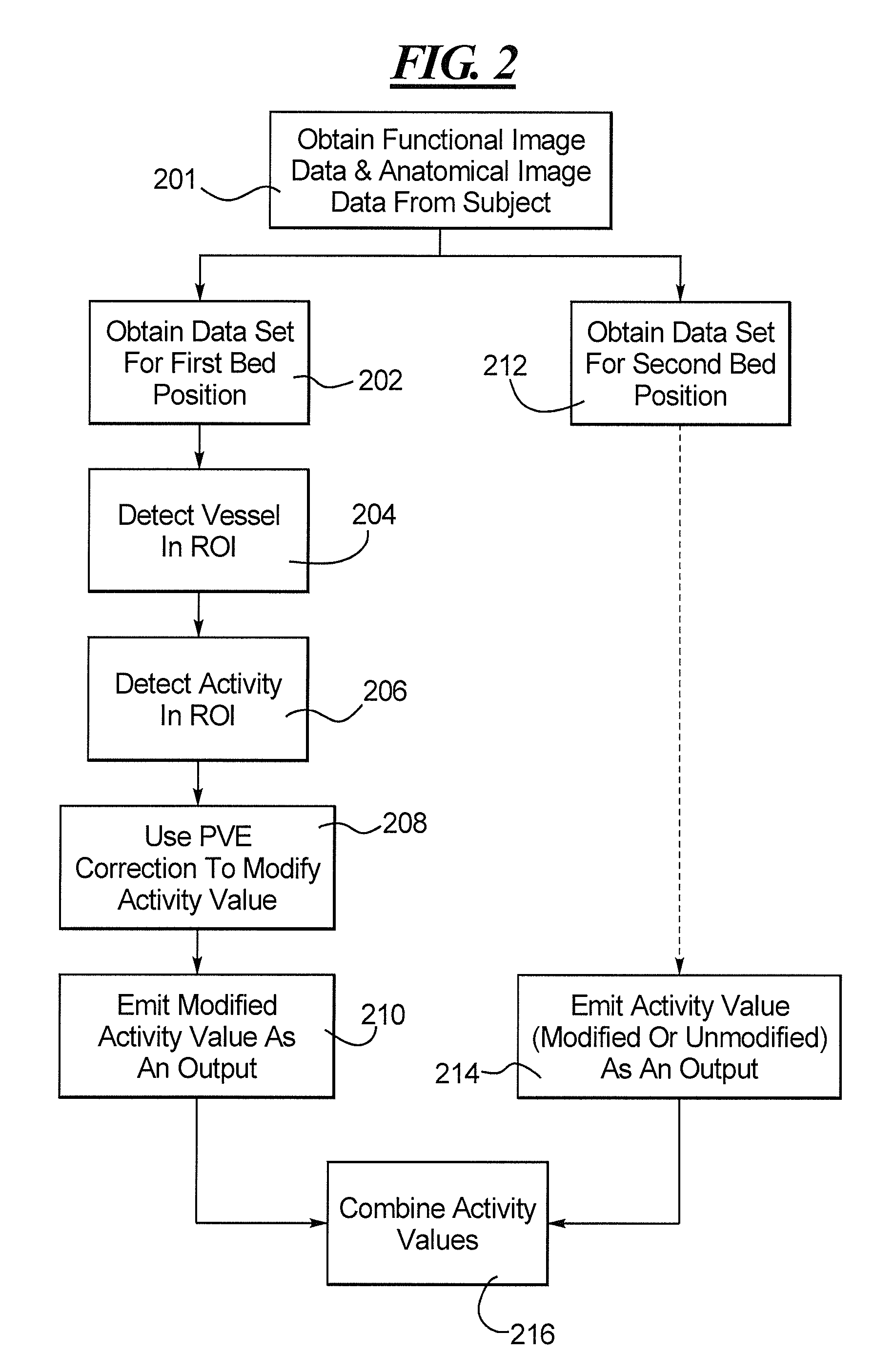
[0042]The problem faced is in replacing the single constant VOI in the traditional dynamic scan with a VOI in each bed position that corresponds to the blood pool, and stitching together the resulting measurements into a single time activity curve (TAC). In addition to the additional effort required to collate the BIF data, the partial volume effect (PVE) will cause measured values to be quantitatively incomparable due to the variation in blood vessel diameter across the bed positions (see FIG. 1).

[0043]FIG. 1 illustrates the effect of PVE on activity concentration (102, 104) measured within blood vessels (...


PUM


Abstract

In a method and apparatus for measuring activity of a tracer in a subject in a medical imaging protocol, comparable features in each of a time series of image data sets of the subject are detected. A first activity value for a first region of interest containing a first of the features in a first image data set is obtained, and a size of the first feature is measured. The first activity value is then modified using the measured size of the first feature. A second activity value for a second region of interest containing a second of the features in a second image data set is obtained. The modified first activity value and the second activity value are then combined to produce a measure of activity of the tracer over the time series.

Description

BACKGROUND OF THE INVENTION[0001]1. Field of the Invention[0002]The present invention is directed to methods and apparatus for measuring activity of a tracer in a subject in a medical imaging protocol.[0003]2. Description of the Prior Art[0004]In the medical imaging field, several imaging schemes are known. For example PET (Positron Emission Tomography) is a method for imaging a subject in 3D using an injected radio-active substance which is processed in the body, typically resulting in an image indicating one or more biological functions. Other such functional imaging modalities are known, such as SPECT.[0005]In such functional images, many important pathologies and anatomical structures appear as very high (or low) intensities. For example, a tumour in an FDG-PET image will often appear as a bright region[0006]Kinetic modelling of the imaging data from dynamic PET acquisitions, for example to model one or more biological functions, requires measurement of a blood input function (B...

Claims


Application Information

Patent Timeline
Patent Type & Authority Applications(United States)
IPC IPC(8): G06K9/46
CPCG06T7/20G06T2207/30096G06T2207/10108G06T2207/10104
Inventor KELLY, MATTHEW DAVIDSADDI, KINDA ANNA
Owner SIEMENS MEDICAL SOLUTIONS USA INC
Why Eureka
Try Eureka