Image diagnosis support apparatus, method and program

a support apparatus and image technology, applied in the field of image diagnosis support apparatus, an image diagnosis method and an image diagnosis program, can solve the problems of inability to flexibly separate and display the anatomical structure, inability to separate a region that was not modeled by a partial model, and inability to properly display an image for the purpose of diagnosis, so as to achieve the desired separation display and easily specify the desired position

Active Publication Date: 2013-03-21
FUJIFILM CORP
View PDF9 Cites 15 Cited by
  • Summary
  • Abstract
  • Description
  • Claims
  • Application Information

AI Technical Summary

Benefits of technology

[0051]When the separation condition further determines a rotation direction and a rotation amount for rotating and displaying the anatomical structures, and the separate image generation means generates a three-dimensional medical image in which the anatomical structures are separated and rotated based on the separation condition, it is possible to rotate an anatomical structure in such a manner that a boundary surface that separates the anatomical structure is displayed on a display screen, or the like. Since it is possible to display the anatomical structure that has been rotated in various directions by an arbitrary rotation amount, it is possible to easily perform desired separate display.
[0052]When the specifying means specifies the specified position by specifying a predetermined tomographic image of a plurality of tomographic images constituting the three-dimensional medical image instead of specifying the specified position in the three-dimensional medical image, it is possible to easily specify a desired specified position based on the position of a desired tomographic image.

Problems solved by technology

Therefore, it has been impossible to flexibly separate and display the anatomical structure to be diagnosed based on a desirable position required for the purpose of diagnosis.
Therefore, in some cases, it was impossible to display an image that was appropriately separated for the purpose of diagnosis, such as diagnosis of an abnormal shadow.
It was impossible to separately display a region that was not modeled by a partial model and the like.
Therefore, especially when plural anatomical structures are present in a three-dimensional image, an operation for separate display was complicated.

Method used


Image

  • Image diagnosis support apparatus, method and program
  • Image diagnosis support apparatus, method and program
  • Image diagnosis support apparatus, method and program

Examples


first embodiment

[0092]Next, configuration related to an image diagnosis support apparatus will be described. In the specification of the present application, the same sign will be assigned to the same part, and explanation about the same part will be omitted.

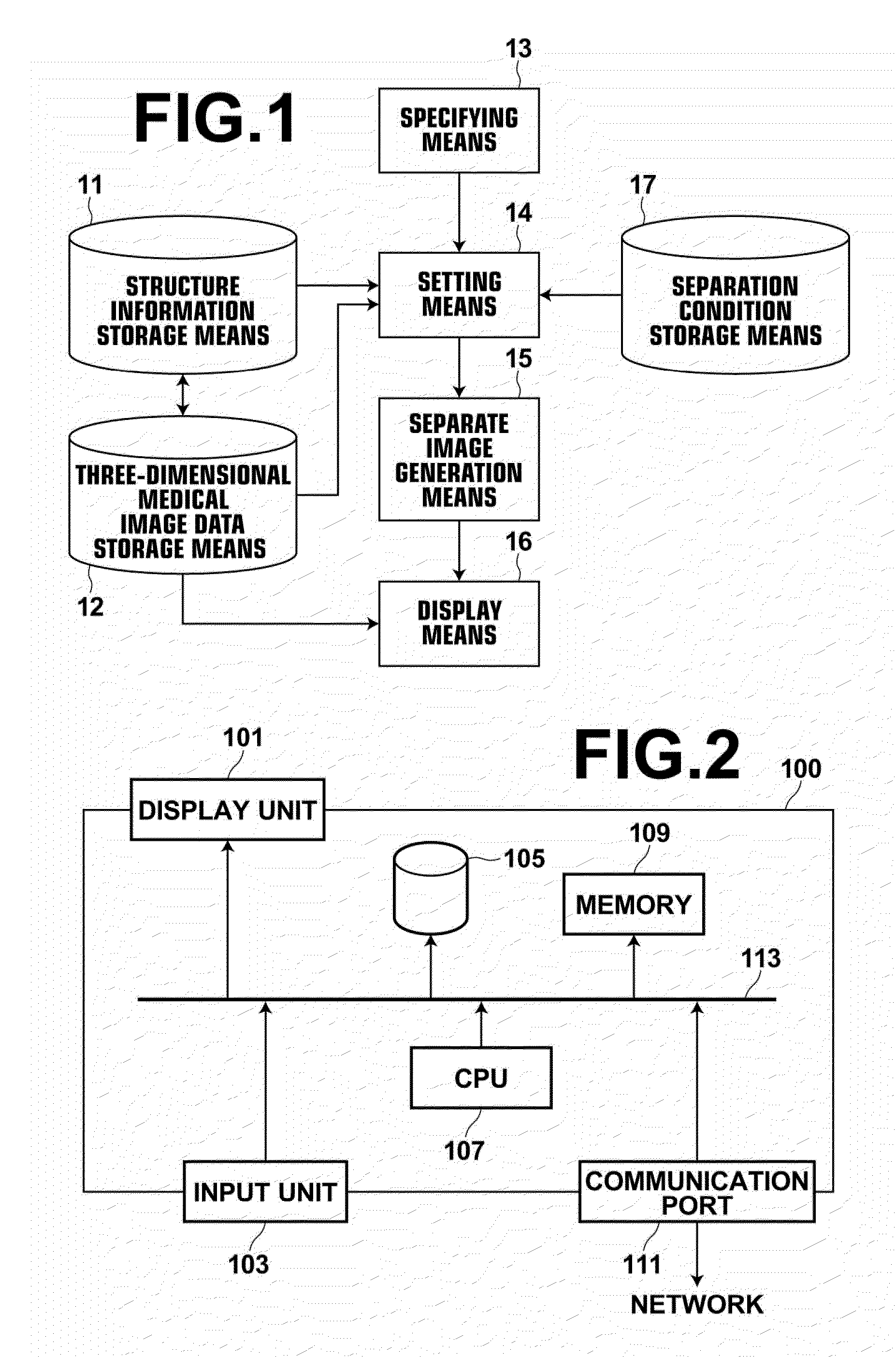
[0093]FIG. 2 is a schematic block diagram illustrating the configuration of an image processing workstation 100. As illustrated in FIG. 2, the image diagnosis support apparatus according to embodiments of the present invention is constituted by the image processing workstation 100 including a display unit 101, such as a liquid crystal monitor, which performs various kinds of display, an input unit 103 including a keyboard, a mouse and the like for performing various kinds of input, a hard disk 105 that stores various kinds of program for controlling the image diagnosis support apparatus according to the embodiments of the present invention and various kinds of data, such as image data, a CPU 107 that controls the image diagnosis support appara...

second embodiment

[0144]FIGS. 9A through 9C are diagrams illustrating examples of hierarchical display of a head according to an embodiment of the present invention. FIG. 9A is an image diagram of a three-dimensional medical image of the head before separate display. FIG. 9B is a diagram illustrating an example of separate display of the head in rank 1 by hierarchical display option. FIG. 9C is a diagram illustrating an example of separate display of the head in rank 2 by hierarchical display option. FIG. 10 is a diagram illustrating a separation condition table 171 including examples of separation conditions with hierarchical display option. FIG. 11 is a flow chart representing a flow of processing of hierarchical display option by an image diagnosis support apparatus of the

[0145]Separate display processing by hierarchical display option will be described based on the flow chart illustrated in FIG. 11. Processing of S201 is similar to processing of S101, and processing of S202 is similar to processi...

third embodiment

[0156]Further, a storage means, such a database, that stores relative arrangement of anatomical structures by using various known methods maybe further provided to set hierarchical display option of the separation condition. The database records relative positions of plural anatomical structures obtained by learning image data including a three-dimensional medical image representing the plural anatomical structures. The hierarchical display option may be automatically set for a boundary surface or a cutting surface for separating an anatomical structure in such a manner that different ranks are sequentially set from the outer-side anatomical structure toward the inner-side anatomical structure. Since it is possible to separately display, step by step, without setting complicated separation condition, it is possible to efficiently support diagnosis using images. In embodiments of the present invention, the separation condition includes partial separation option in which separate disp...


PUM


Abstract

At least one specified position and, if necessary, a cutting surface are specified in a three-dimensional medical image. Plural anatomical structures present within a predetermined range from the specified position are extracted, as structures to be separated, by referring to a structure information storage unit that stores plural anatomical structures and a separation condition storage unit that stores a separation condition for each anatomical structure of a subject to determine, based on the specified position, a boundary surface and, if necessary, a cutting surface for separately displaying the plural anatomical structures. The boundary surface corresponding to the structures to be separated and the specified position and, if necessary, the cutting surface are set based on the separation condition. A three-dimensional medical image in which the structures to be separated are separated by the boundary surface and, if necessary, by the cutting surface is generated, and displayed.

Description

BACKGROUND OF THE INVENTION[0001]1. Field of the Invention[0002]The present invention relates to an image diagnosis support apparatus, an image diagnosis method and an image diagnosis program in which a three-dimensional medical image is displayed to support diagnosis.[0003]2. Description of the Related Art[0004]In recent years, medical image display techniques advanced in medical fields. In the medical image display techniques, a three-dimensional medical image reconstructed from plural sets of imaging image data obtained by imaging by an ultrasound diagnosis apparatus, a CT apparatus or an MRI apparatus, and a medical imaging apparatus, such as a PET, is displayed to support image diagnosis performed in medical fields. In such image diagnosis support techniques, a technique for visualizing a detail of a desired region in a three-dimensional medical image to satisfy the purpose of diagnosis has been drawing attention.[0005]For example, Japanese Unexamined Patent Publication No.[000...

Claims


Application Information

Patent Timeline
Patent Type & Authority Applications(United States)
IPC IPC(8): G06K9/00
CPCG06F19/321G06K9/00201G06T2219/2004G06T2219/008G06T19/20G16H30/40G16H30/20G16H50/20G06V20/64
Inventor SHIRASAKA, HAJIMENAKAMURA, KEIGO
Owner FUJIFILM CORP
Why Eureka
Try Eureka