Methods of detecting cancer

a detection method and cancer technology, applied in the field of molecular classification of diseases, can solve the problems of poor prognosis, major health challenges of cancer, and most vexing aspects of cancer remain early detection

Inactive Publication Date: 2013-06-06
MYRIAD GENETICS
View PDF12 Cites 76 Cited by
  • Summary
  • Abstract
  • Description
  • Claims
  • Application Information

AI Technical Summary

Benefits of technology

[0004]Mutations in certain genes are associated with cancer in general and with specific cancer types. For example, inactivating mutations in the TP53 gene are found in approximately 50% of all solid tumors and activating mutations in the KRAS or BRAF genes are often found in colorectal cancer. It has been discovered that screening patients for mutations in certain genes can detect and classify cancer. More specifically, it has been determined that (a) screening certain genes (e.g., APC, EGFR, KRAS, PTEN, and TP53) for mutations will detect nearly 95% of all cancers, while (b) screening certain genes (e.g., AIM1, APC, CDKN2A, EGFR, FBN2, FBXW7, FLJ13479, IDH1, KRAS, PIK3CA, PIK3R1, PTEN, RB1, SMAD4, TGFBR2, TNN, and TP53) for mutations can accurately classify the cancer (e.g., as breast cancer, colon cancer, glioblastoma, pancreatic cancer, etc.).

Problems solved by technology

Cancer is a major health challenge.
Despite recent advances in molecular and imaging diagnostics, one of the most vexing aspects of cancer remains early detection.
In fact, for certain types of cancer—e.g., pancreatic adenocarcinoma—detection often occurs so late as to practically preclude any good prognosis.

Method used


Image

  • Methods of detecting cancer
  • Methods of detecting cancer
  • Methods of detecting cancer

Examples


example 1

Using Somatic Mutations to Determine Tumor Site

Methods

[0103]Consider a sample from a patient with some type of cancer. The mutation screening of this sample identifies somatic mutations in n genes g1, g2, . . . , gn. Assuming that somatic mutations occur independently, the probability that this patient has cancer of type c is given by the following equation:

P(c|g1,g2, . . . ,gn)=P0(c)ΠiM(gi|c) / ΣtP0(t)ΠiM(gi|t)  (1)

where the product is taken over all genes mutated in the sample (i=1, 2, . . . , n) and the sum is taken over all cancer types t. M(g|c) is the frequency of somatic mutations in gene g in cancer type c. See FIG. 3 (with mutation frequencies based on data from COSMIC [Catalogue of Somatic Mutations in Cancer] database). P0(c) is the a priori probability of cancer type c given that the patient has a cancer. See FIG. 4 (with these a priori probabilities based on cancer incidences published by the American Cancer Society). It should be noted that for some cancers (such as ovar...

example 2

Using Somatic Mutations to Detect Presence of Cancer

Method

[0122]Since somatic mutations are very specific to cancer or pre-cancerous conditions, the main performance measure of using mutation screening of a set of genes is its sensitivity. The sensitivity of screening for any cancer depends on sensitivities within individual cancers as well as on the incidences of the cancers. The sensitivity was defined by the following equation:

S=ΣtP0(t)S(t)  (4)

where S(t) was the sensitivity within cancer type t. S(t) was defined as the percentage of patients with somatic mutations in one or more genes within a predefined set of one or more genes.

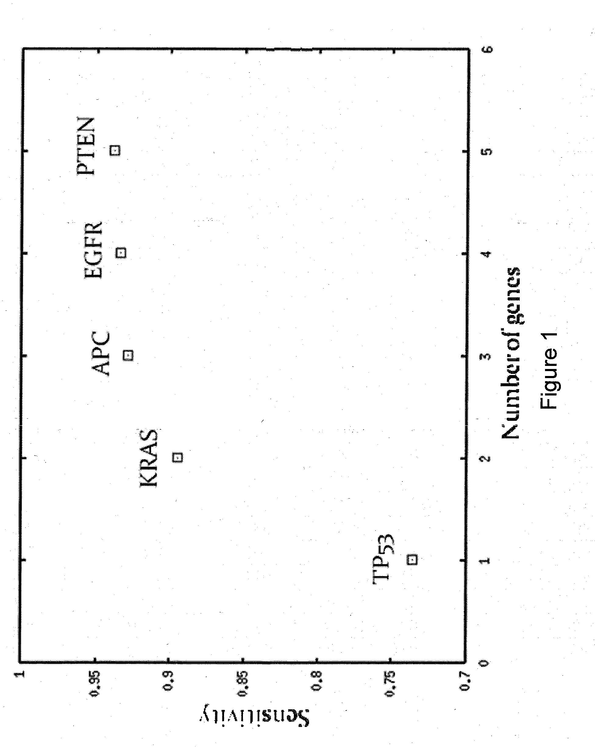
[0123]The following algorithm was used to define a small set of genes with high sensitivity:[0124]1. Started with all available samples and an empty list of genes.[0125]2. Within current set of samples, found the gene with highest sensitivity calculated according to Equation (4). This gene was added to the list of genes.[0126]3. Repeated Steps 1 & 2 unti...

example 3

Detecting Mutations in Exosomes

Method

[0134]To confirm our ability to detect cancer-related mutations in serum exosomes, cell culture supernatants (1-10 ml from ovarian and colon cancer cell lines) or ovarian and colon cancer patient serum samples (1-3 ml) were used to prepare exosomes by high-speed centrifugation. Total RNA was extracted from exosomal pellets and converted to cDNA by standard methods. PCR amplicons for a set of mutation hot spots in TP53, KRAS, EGFR and APC were designed and optimized for multiplexing. Exosomal cDNA was pre-amplified with a multiplex of all amplicons. The pre-amplification product was split into separate reactions and re-amplified with the individual target amplicons. Re-amplification primers were synthesized with tails for dye-primer sequencing. Individual PCR products were sequenced by dye-primer chemistry to identify particular mutations.

Results

[0135]Mutations were found in exosomes harvested from cell lines as follows:

TABLE 5ExosomalExosomalCell...


PUM

PropertyMeasurementUnit
frequencyaaaaaaaaaa
thresholdaaaaaaaaaa

Abstract

Methods and compositions involving molecular markers for the detection and characterization of cancer in a patient are provided.

Description

CROSS REFERENCE TO RELATED APPLICATIONS[0001]This application claims the priority benefit of PCT / US10 / 037,659, filed Jun. 7, 2010, which claims the priority benefit of U.S. Provisional Application Ser. No. 61 / 184,685 (filed on Jun. 5, 2009), which is hereby incorporated by reference in its entirety.FIELD OF THE INVENTION[0002]The invention generally relates to a molecular classification of disease and particularly to molecular markers for cancer and methods of use thereof.BACKGROUND OF THE INVENTION[0003]Cancer is a major health challenge. Nearly 560,000 people die from cancer annually in the United States alone, representing almost 23% of all deaths. Despite recent advances in molecular and imaging diagnostics, one of the most vexing aspects of cancer remains early detection. In fact, for certain types of cancer—e.g., pancreatic adenocarcinoma—detection often occurs so late as to practically preclude any good prognosis. Thus there is an urgent need for sensitive methods of detectin...

Claims


Application Information

Patent Timeline
Patent Type & Authority Applications(United States)
IPC IPC(8): C40B30/00C40B40/08
CPCC12Q2600/156C12Q1/6886G16H50/20
Inventor GUTIN, ALEXANDERLANCHBURY, JERRYWAGNER, SUSANNEABKEVICH, VICTOR
Owner MYRIAD GENETICS
Why Eureka
Try Eureka