
Method of classifying counting leucocytes

a technology of leukocytes and counting methods, applied in the field of classifying and counting leukocytes, can solve the problems of complex separation of mononuclear leukocytes by specific gravity centrifugation, inability to precisely detect and classify immature leukocytes, and the inability to differentiate leukocytes

Active Publication Date: 2005-09-15
SYSMEX CORP
View PDF3 Cites 62 Cited by
  • Summary
  • Abstract
  • Description
  • Claims
  • Application Information

AI Technical Summary

Benefits of technology

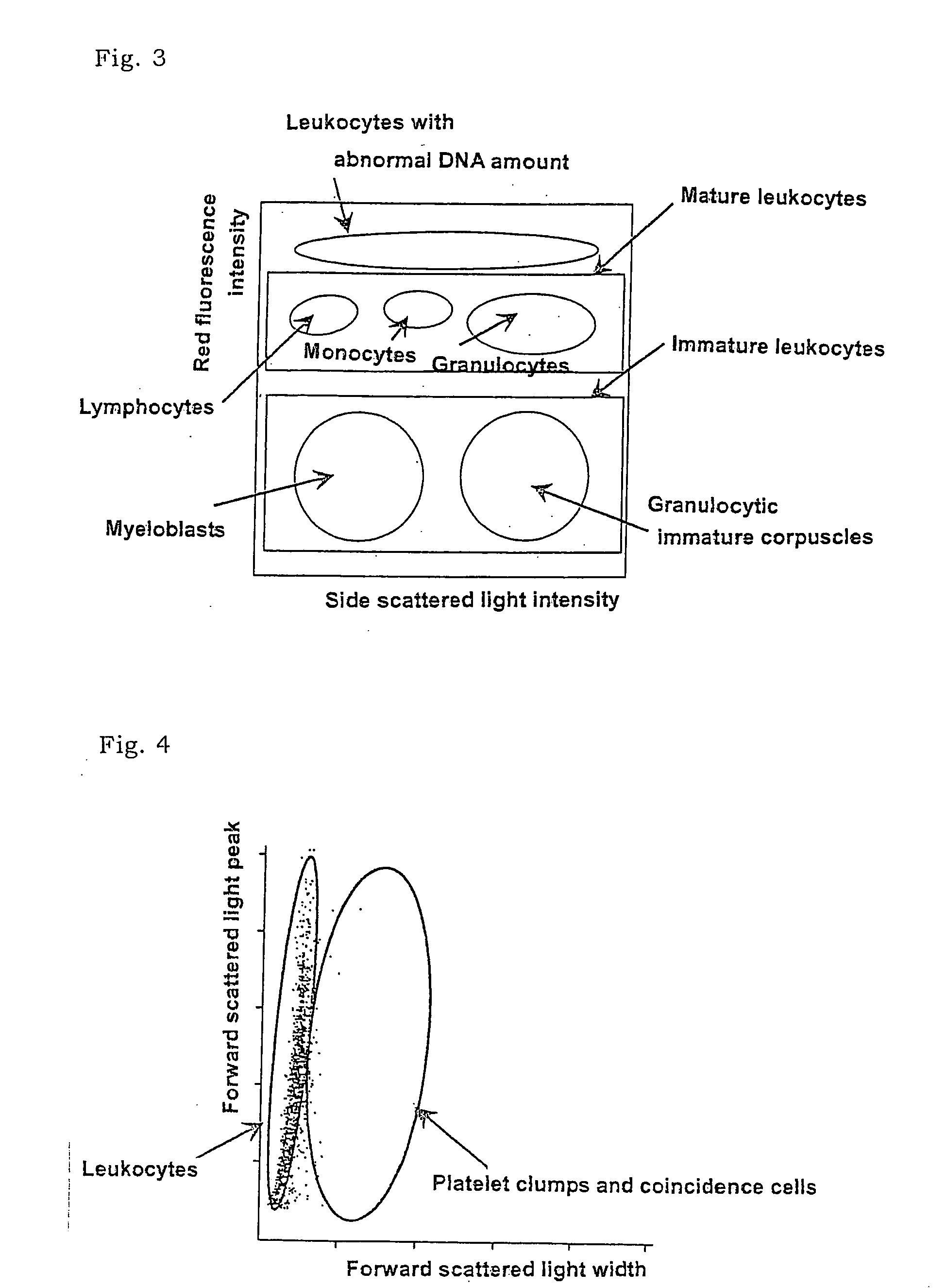
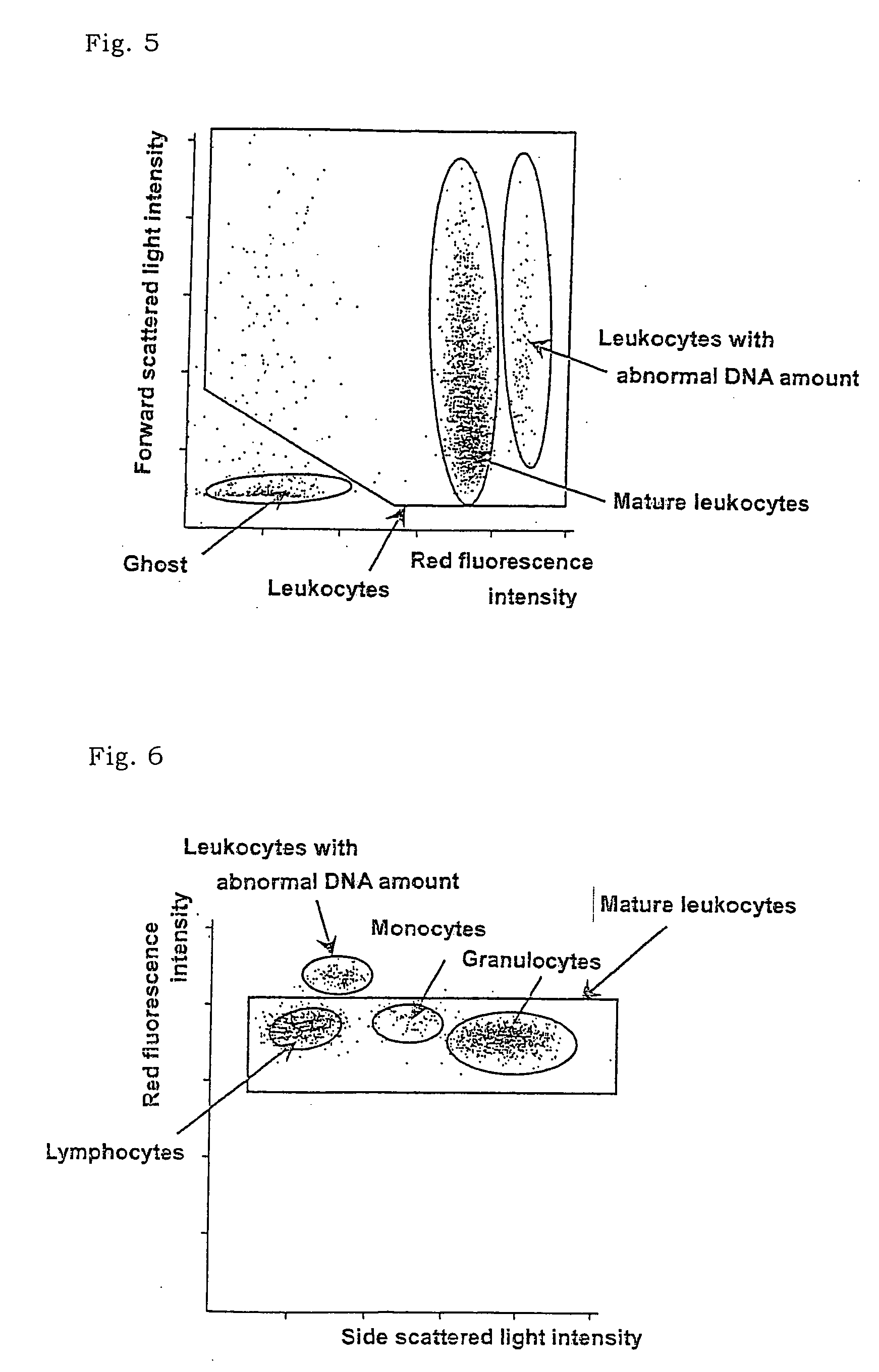
[0007] According to the present invention, there is provided a method which makes it possible to classify and count leukocytes with abnormal DNA amount and, simultaneously, immature leukocytes and mature leukocytes, by maintaining immature leukocytes in a viable state, treating other leukocytes with a hemolytic agent which damages other leukocytes, staining the damaged cells with a fluorescent dye which can stain damaged cells, measuring scattered light and fluorescence of the resulting blood corpuscles, and using the difference in the intensity of the scattered light peak and the difference in the scattered light width.
[0061] The light source of flow cytometer is not particularly limited, and is selected from the sources having a suitable wavelength for excitation of the dye. For example, argon laser, He—Ne laser, red semiconductor laser, blue semiconductor laser, and the like may be used. In particular, a semiconductor laser is much cheaper than gas laser, thus cost for an apparatus can be reduced drastically.
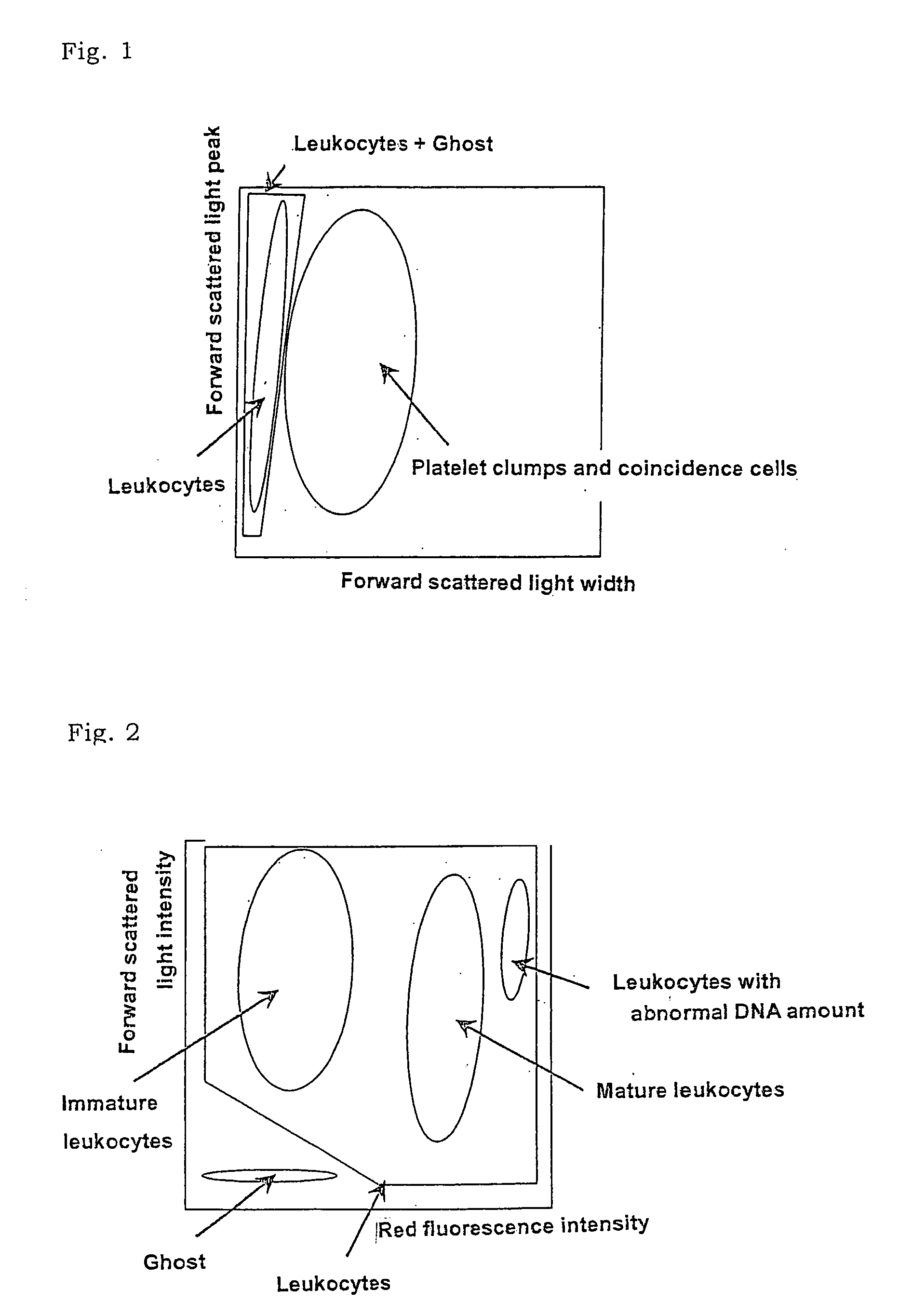
[0062] In the step (3), platelet clumps / coincidence cells and leukocytes are classified using the difference in the intensity of the scattered light peak and the difference in the scattered light width. Specifically, for example, a scattergram is prepared in which the X-axis indicates the forward scattered light width and the Y-axis indicates the forward scattered light peak. As shown in FIG. 1, for example, platelet clumps and coincidence cells as well as leukocytes and ghost form respective groups which distribute in the scattergram. From all of the cells on the scattergram, the platelet clumps and coincidence cells are removed to classify the platelet clumps / coincidence cells and the leukocytes / ghost. By this operation, it is possible to prevent an appearance of platelet clumps and coincidence cells in the area of the leukocytes with abnormal DNA amount, and to precisely classify and count leukocytes with abnormal DNA amount.

Problems solved by technology

Separation of the mononuclear leukocytes by specific gravity centrifugation is complicated in operation and requires 1 hour or more for completion of the separation.
Although normal leukocytes can be highly precisely classified and counted with these cytometers, immature leukocytes can not precisely be detected and classified because the cytometers are greatly influenced by platelet clumps and coincidence cells.
In this method, however, it was not possible to differentiate leukocytes with abnormal DNA amount from platelet clumps and coincidence cells, thus it was difficult to precisely classify and count leukocytes with abnormal DNA amount.

Method used


Image

  • Method of classifying counting leucocytes
  • Method of classifying counting leucocytes
  • Method of classifying counting leucocytes

Examples


example 1

[0088] A reagent comprising an aqueous solution of the following composition was prepared.

(The present method)Polyoxyethylene(16)oleyl ether24.0gSodium N-lauroylsarcosinate1.5gDL-Methionine20.0g1N-NaOH0.3gNaCl4.0gDye of formula (VI)3.0mgHEPES12.0gPure water1000ml

[0089] The above-mentioned reagent (1 ml) was mixed with 33 μl of blood collected from a patient suffering from lymphoid leukemia, and after a lapse of 10 seconds, forward low angle scattered light, side scattered light, and red fluorescence were measured with a flow cytometer (light source: red semiconductor laser, wavelength: 633 nm).

(A standard method for measurement of the DNA amount)Trisodium citrate100mgTriton X-100 (Wako Pure Chemical Industries, Ltd.)0.2gPropidium iodide (Sigma)0.2gRO water100ml

[0090] The above-mentioned reagent (1 ml) was mixed with 100μl of blood collected from the above-mentioned patient, and after a lapse of 30 minutes, red fluorescence was measured with a flow cytometer (light source: argon ...

example 2

[0094] A reagent comprising an aqueous solution of the following composition was prepared.

(The present method)Polyoxyethylene(16)oleyl ether24.0gSodium N-lauroylsarcosinate1.5gDL-Methionine20.0g1N-NaOH0.3gNaCl4.0gDye of formula (VII)3.0mgHEPES12.0gPure water1000ml

[0095] The above-mentioned reagent (1 ml) was mixed with 33 μl of blood collected from a patient suffering from acute myelocytic leukemia (AML), and after a lapse of 10 seconds, forward low angle scattered light, side scattered light, and red fluorescence were measured with a flow cytometer (light source: red semiconductor laser, wavelength: 633 nm).

(A standard method for measurement of the DNA amount)Trisodium citrate100mgTriton X-100 (Wako Pure Chemical Industries, Ltd.)0.2gPropidium iodide (Sigma)0.2gRO water100ml

[0096] The above-mentioned reagent (1 ml) was mixed with 100 μl of blood collected from the above-mentioned patient, and after a lapse of 30 minutes, red fluorescence was measured with a flow cytometer (ligh...

example 3

[0100] The reagents with the same composition as in Example 1 were used. The reagent of the present method (1 ml) was mixed with 33 μl of bone marrow fluid collected from a patient suffering from osteomyelodysplasia syndrome, and after a lapse of 7 seconds and 13 seconds, forward low angle scattered light, side scattered light, and red fluorescence were measured with a flow cytometer (light source: red semiconductor laser, wavelength: 633 nm).

(A standard Method for Measurement of the DNA Amount)

[0101] The reagent (1 ml) for the standard method for measurement of the DNA amount was mixed with 100 μl of bone marrow fluid collected from the above-mentioned patient, and after a lapse of 30 minutes, red fluorescence was measured with a flow cytometer (light source: argon ion laser, wavelength: 4-88 nm).

[0102] The results are shown in a scattergram (FIG. 11) for the case of 7 seconds of the reaction time in which the X-axis indicates the forward low angle scattered light width and the...


PUM

PropertyMeasurementUnit
temperatureaaaaaaaaaa
receiving angleaaaaaaaaaa

Abstract

There is provided a method for classifying and counting leukocytes with abnormal DNA amount, which comprises: (1) a step of staining cells in a sample obtained from a hematological sample by treatment with a hemolytic agent to lyse erythrocytes, with a fluorescent dye which can make a difference in the fluorescence intensity at least among mature leukocytes, leukocytes with abnormal DNA amount and immature leukocytes; (2) a step of introducing the sample containing the stained cells into a flow cytometer to measure scattered light and fluorescence of the respective cells; (3) a step of classifying leukocytes and coincidence cells / platelet clumps utilizing a difference in the intensity of a scattered light peak and a difference in the scattered light width; (4) a step of classifying and counting mature leukocytes, leukocytes with abnormal DNA amount and immature leukocytes, utilizing a difference in the scattered light intensity and a difference in the fluorescence intensity of leukocytes classified in the step (3).

Description

FIELD OF THE INVENTION [0001] The present invention relates to a method for classifying and counting leukocytes. More particularly, it relates to a method for classifying and counting leukocytes, particularly leukocytes with abnormal DNA amount, in hematological samples utilizing a flow cytometer. BACKGROUND ART [0002] In the field of clinical laboratory test, classifying and counting leukocytes with abnormal DNA amount or immature leukocytes provide very useful information in diagnosis of diseases. For example, leukocytes in normal peripheral blood generally consist of these five-types, i.e., lymphocytes, monocytes, basophiles, eosinophils and neutrophils, and the DNA amount of leukocytes is constant. However, leukocytes with abnormal DNA amount appear, for example, in blood disorders such as viral diseases or lymphocytic leukemia, and immature leukocytes appear in such blood disorders as myclocytic leukemia. [0003] Usually, in order to measure leukocytes with abnormal DNA amount, ...

Claims


Application Information

Patent Timeline
Patent Type & Authority Applications(United States)
IPC IPC(8): G01N15/00G01N15/14G01N33/52
CPCG01N2015/1477G01N33/52
Inventor TSUJI, TOMOHIROMIZUKAMI, TOSHIHIROKONISHI, AYAMORI, YUSUKENAKAZAWA, YUKIE
Owner SYSMEX CORP
Learn More