Ultrasound imaging system and method

a technology of ultrasonic imaging and ultrasonic sonic, applied in ultrasonic/sonic/infrasonic diagnostics, instruments, tomography, etc., can solve the problems of easy disorientation of users within the volume, difficult for even an experienced clinician to remain oriented with respect to the patient's anatomy,

Inactive Publication Date: 2013-06-13
GENERAL ELECTRIC CO
View PDF3 Cites 30 Cited by
  • Summary
  • Abstract
  • Description
  • Claims
  • Application Information

AI Technical Summary

Problems solved by technology

Due to all of the image manipulations that are possible on conventional ultrasound imaging systems, it is easy for users to become disoriented within the volume.
Between adjustments and rotations to volume-rendered images and adjustments, including translations, rotations, and tilts to the planar images, it may be difficult for even an experienced clinician to remain oriented with respect to the patient's anatomy while manipulating and adjusting the volume-rendered image and / or the planar images.

Method used


Image

  • Ultrasound imaging system and method
  • Ultrasound imaging system and method
  • Ultrasound imaging system and method

Examples


Embodiment Construction

[0014]In the following detailed description, reference is made to the accompanying drawings that form a part hereof, and in which is shown by way of illustration specific embodiments that may be practiced. These embodiments are described in sufficient detail to enable those skilled in the art to practice the embodiments, and it is to be understood that other embodiments may be utilized and that logical, mechanical, electrical and other changes may be made without departing from the scope of the embodiments. The following detailed description is, therefore, not to be taken as limiting the scope of the invention.

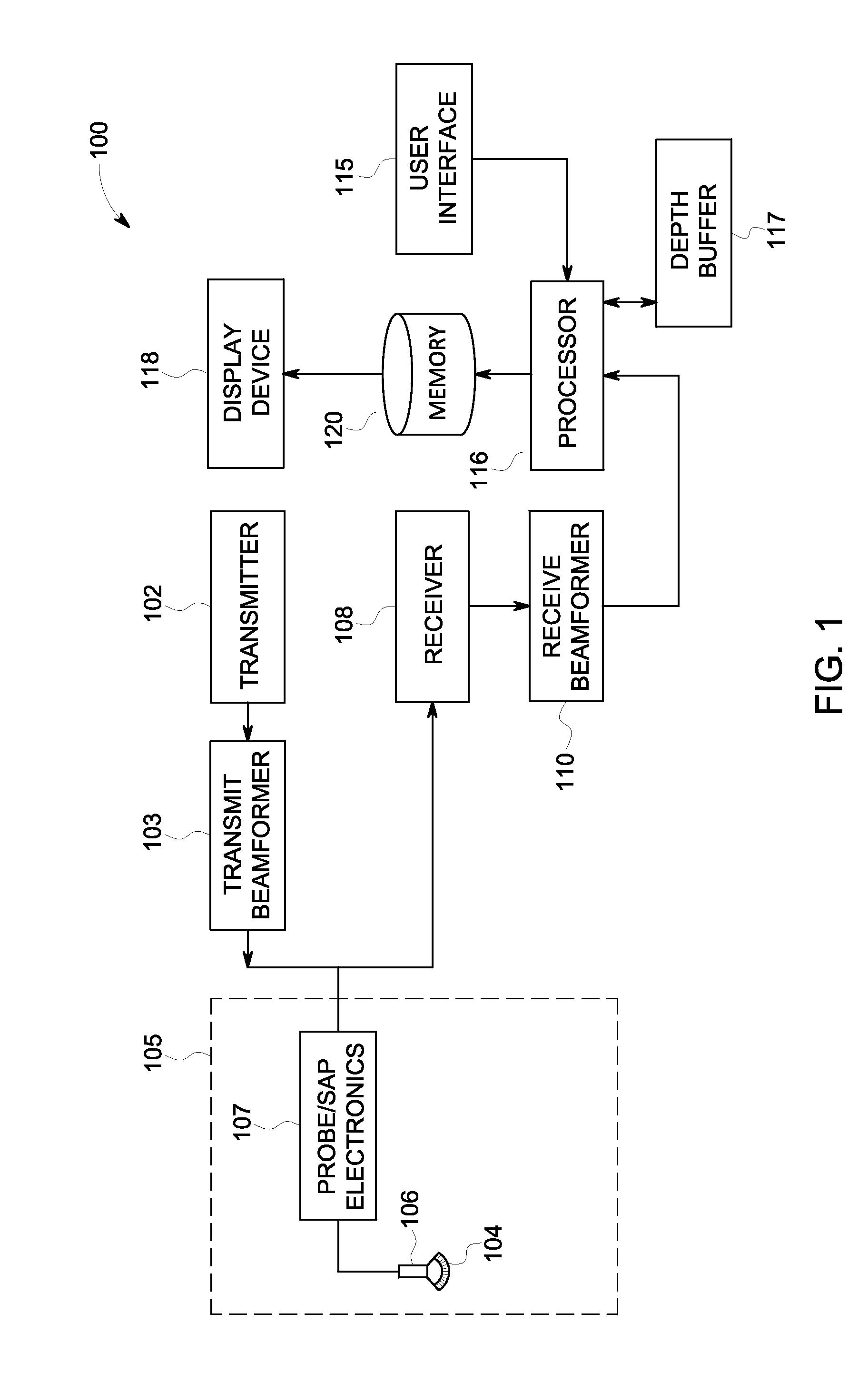
[0015]FIG. 1 is a schematic diagram of an ultrasound imaging system 100 in accordance with an embodiment. The ultrasound imaging system 100 includes a transmitter 102 that transmits a signal to a transmit beamformer 103 which in turn drives transducer elements 104 within a transducer array 106 to emit pulsed ultrasonic signals into a structure, such as a patient (not shown). A...


PUM


Abstract

An ultrasound imaging system and method for ultrasound imaging. The method includes generating a volume-rendered image from three-dimensional ultrasound data. The volume-rendered image is colorized with at least two colors according to a depth-dependent color scheme. The method includes displaying the volume-rendered image. The method includes generating a planar image from the three-dimensional ultrasound data, where the planar image is colorized according to the same depth-dependent color scheme. The method includes displaying the planar image.

Description

FIELD OF THE INVENTION[0001]This disclosure relates generally to an ultrasound imaging system and method for displaying a volume-rendered image and an planar image that are both colorized according to the same depth-dependent scheme.BACKGROUND OF THE INVENTION[0002]Conventional ultrasound imaging systems acquire three-dimensional ultrasound data from a patient and are then able to generate and display multiple types of images from the three-dimensional ultrasound data. For example, conventional ultrasound imaging systems may generate and display a volume-rendered image based on the three-dimensional ultrasound data and / or conventional ultrasound imaging systems may generate one or more planar images from the three-dimensional ultrasound data. The volume-rendered image is a perspective view of surfaces rendered from the three-dimensional ultrasound data while the planar image is an image of a plane through the volume included within the three-dimensional ultrasound data. Users would ...

Claims


Application Information

Patent Timeline
Patent Type & Authority Applications(United States)
IPC IPC(8): A61B8/13
CPCG06T15/08G06T19/00G06T2219/008G06T2219/028G01S7/52074A61B8/466A61B8/483G01S15/8993G01S7/52071G06T2219/2012
Inventor ORDERUD, FREDRIK
Owner GENERAL ELECTRIC CO
Why Eureka
Try Eureka