
Device with Biological Tissue Scaffold for Percutaneous Closure of an Intracardiac Defect and Methods Thereof

a biological tissue and intracardiac defect technology, applied in the field of devices, can solve the problems of devices suffering, particularly worrisome patients, and heart failure, and achieve the effect of reducing or minimizing the size of the devi

Active Publication Date: 2013-09-26
WL GORE & ASSOC INC
View PDF5 Cites 0 Cited by
  • Summary
  • Abstract
  • Description
  • Claims
  • Application Information

AI Technical Summary

Benefits of technology

The present invention provides a device to block heart defects. This device is different from previous ones because it uses a biological tissue scaffold made from collagen. This scaffold is made from a specific type of biological tissue from the small intestine of a pig. This new design solves the problems associated with previous devices, making it a better choice for heart defect repair.

Problems solved by technology

The deformities result from the incomplete formation of the septum, or muscular wall, between the chambers of the heart and can cause significant problems.
Ultimately, the deformities add strain on the heart, which may result in heart failure if they are not corrected.
This is particularly worrisome to patients who are prone to forming venous thrombus, such as those with deep vein thrombosis or clotting abnormalities.
Such devices suffer, however, from several disadvantages, including thrombus formation, chronic inflammation, and residual leaks.

Method used


Image

  • Device with Biological Tissue Scaffold for Percutaneous Closure of an Intracardiac Defect and Methods Thereof
  • Device with Biological Tissue Scaffold for Percutaneous Closure of an Intracardiac Defect and Methods Thereof
  • Device with Biological Tissue Scaffold for Percutaneous Closure of an Intracardiac Defect and Methods Thereof

Examples


Embodiment Construction

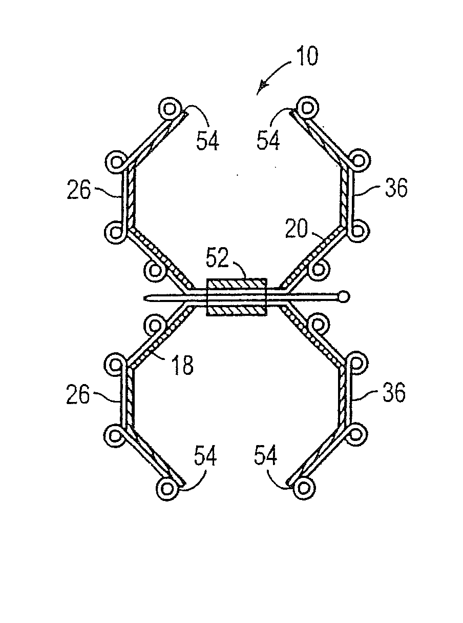
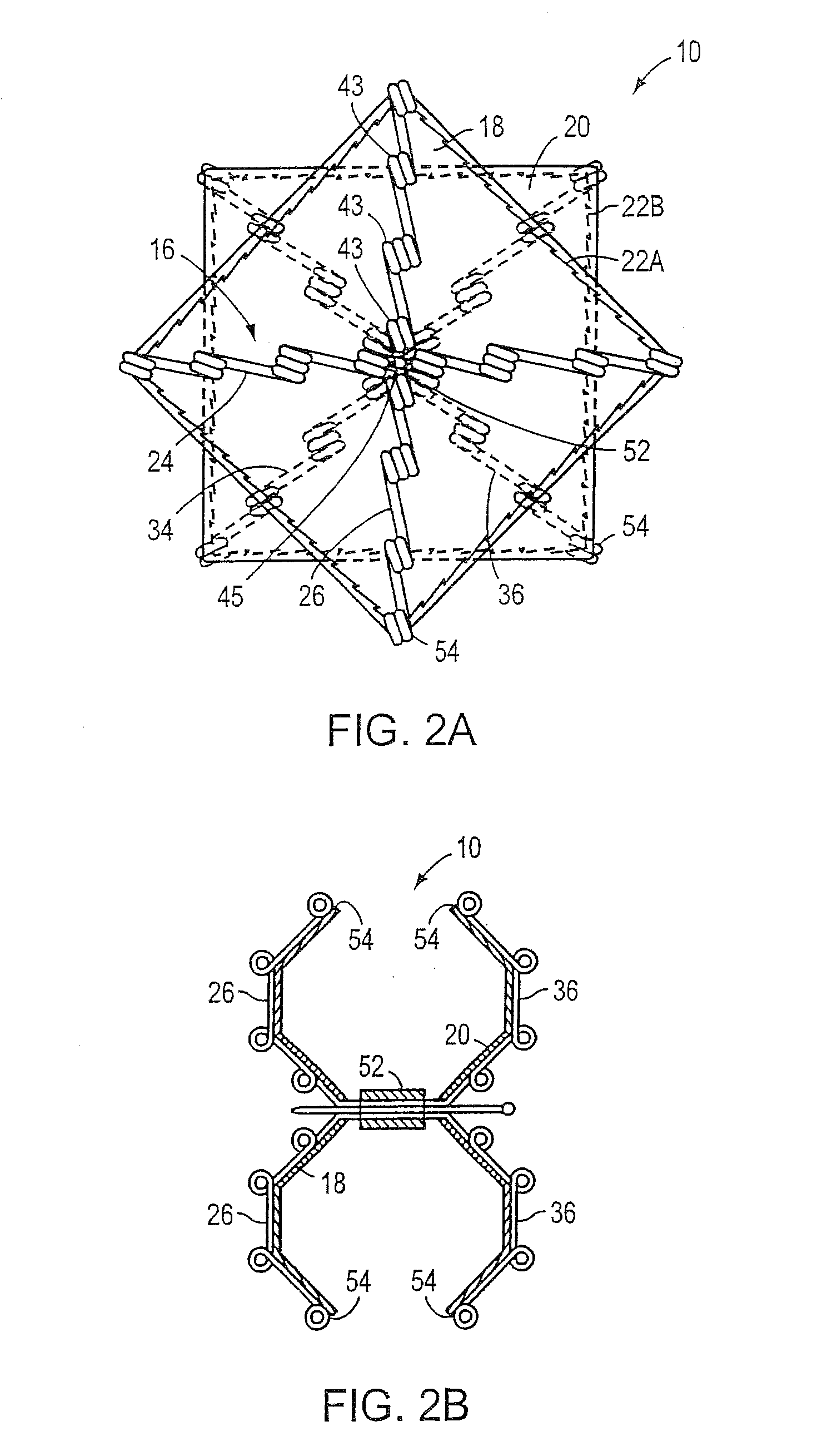
[0028]The present invention provides an intracardiac occluder for the repair of intracardiac defects, such as, for example, a patent foramen ovale, an atrial septal defect, a ventricular septal defect, and left atrial appendages. The intracardiac occluder includes a structural framework and a biological tissue scaffold adhered thereto.

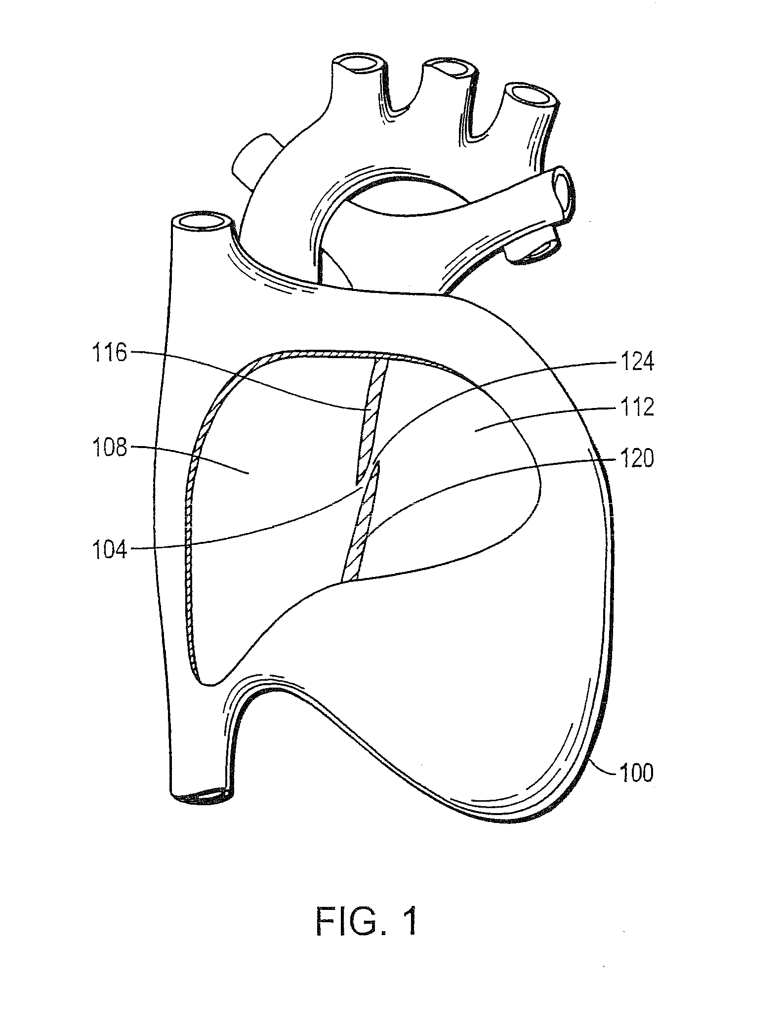
[0029]FIG. 1 depicts a cutaway view of a heart 100. The heart 100 includes a septum 104 that divides a right atrium 108 from a left atrium 112. The septum 104 includes a septum primum 116, a septum secundum 120, and an exemplary intracardiac defect 124, which is to be corrected by the intracardiac occluder of the present invention, between the septum primum 116 and the septum secundum 120. Specifically, a patent foramen ovale 124 is shown as an opening through the septum 104. The patent foramen ovale 124 provides an undesirable fluid communication between the right atrium 108 and the left atrium 112. Under certain conditions, a large patent foramen ova...


PUM

PropertyMeasurementUnit
lengthaaaaaaaaaa
bioresorbableaaaaaaaaaa
corrosion resistantaaaaaaaaaa

Abstract

The invention provides an intracardiac occluder, which has biological tissue scaffolds as occlusion shells, for the percutaneous transluminal treatment of an intracardiac defect. The intracardiac occluder includes a proximal support structure supporting the proximal occlusion shell and a distal support structure supporting the distal occlusion shell. In one embodiment, biological tissue derived from the tunica submucosa layer of the porcine small intestine forms the occlusion shells.

Description

CROSS-REFERENCE TO RELATED APPLICATIONS[0001]This application is a divisional application of U.S. application Ser. No. 11 / 705,380 filed Feb. 12, 2007, now pending; which is a continuation application of U.S. application Ser. No. 10 / 453,709 filed Jun. 3, 2003, now abandoned; which claims the benefit under 35 USC §119(e) to U.S. Application Ser. No. 60 / 385,274 filed Jun. 3, 2002, now expired. The disclosure of each of the prior applications is considered part of and is incorporated by reference in the disclosure of this application.BACKGROUND OF THE INVENTION[0002]1. Field of the Invention[0003]The invention generally relates to devices and related methods for treating intracardiac defects. More particularly, the invention provides an intracardiac occluder with a biological tissue scaffold, and related methods, for the percutaneous closure of intracardiac defects.[0004]2. Background Information[0005]The human heart is divided into four compartments or chambers. The left and right atri...

Claims


Application Information

Patent Timeline
Patent Type & Authority Applications(United States)
IPC IPC(8): A61B17/00A61B17/08A61B17/12
CPCA61B17/0057A61B17/12122A61B17/12172A61F2310/00365A61B2017/00592A61B2017/00606A61B2017/1205A61B2017/00575
Inventor DEVELLIAN, CAROL A.CARR, ROBERT M.
Owner WL GORE & ASSOC INC
Learn More