
Dynamic MR Imaging of Patients with Breast Cancer - Establishment and Comparison of Different Analytical Methods for Tissue Perfusion and Capillary Permeability

a technology of mr imaging and dynamic imaging, which is applied in the direction of magnetic variable regulation, sensors, diagnostics, etc., can solve the problems of low specificity, limited diagnostic performance of radiological image modalities, and difficult to distinguish benign and malignant tumors by contrast-enhanced mr imaging, so as to improve the probability of movement and reduce the effect of movemen

Inactive Publication Date: 2014-02-06
SUNNMORE MR KLINIKK
View PDF0 Cites 22 Cited by
  • Summary
  • Abstract
  • Description
  • Claims
  • Application Information

AI Technical Summary

Benefits of technology

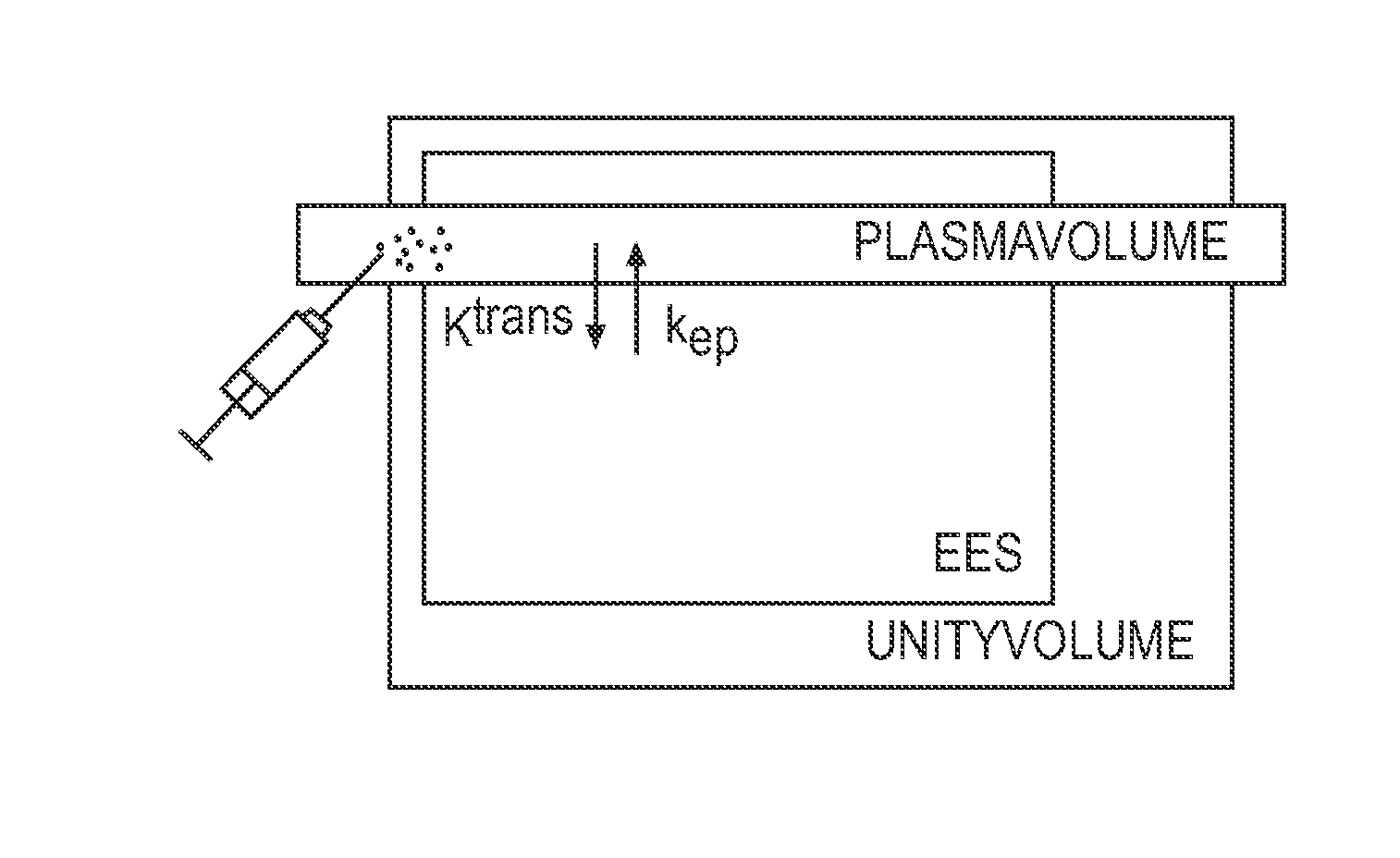
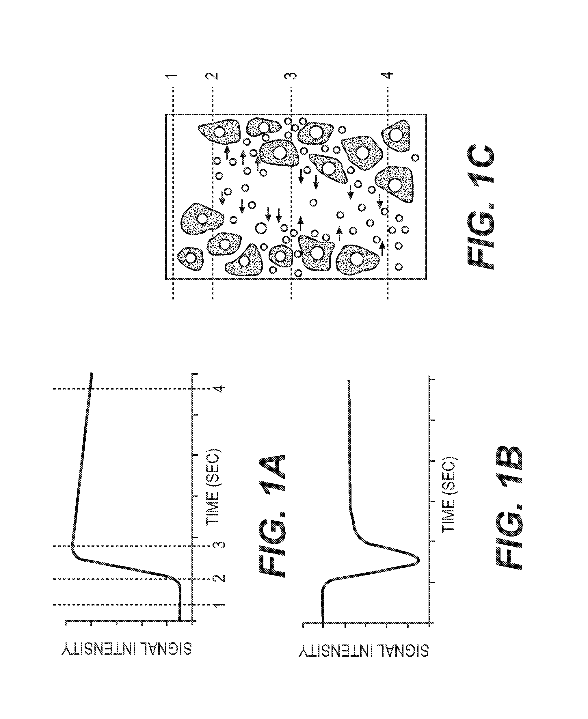
The patent text describes a method for improving the diagnosis of breast lesions by analyzing dynamic images and using a contrast substance to enhance the MRI signals. The method involves collecting data from two distinct MRI sequences, one before and one after the contrast substance is injected. The data is then used to calculate a normalized Ktrans, which is a biomarker that can differentiate between benign and malignant breast lesions. The use of the contrast substance helps to reduce errors in the analysis and improve the accuracy of the diagnosis. The method can also help to identify specific types of breast cancer and improve the performance of conventional methods.

Problems solved by technology

However, these radiological image modalities demonstrate limitations with respect to the diagnostic performance and show a generally low specificity, particularly when evaluating young women.
Some of the disadvantages of the prior art has, however, been that it has proven to be difficult to distinguish between benign and malign tumors by contrast-enhanced MR imaging.

Method used


Image

  • Dynamic MR Imaging of Patients with Breast Cancer - Establishment and Comparison of Different Analytical Methods for Tissue Perfusion and Capillary Permeability
  • Dynamic MR Imaging of Patients with Breast Cancer - Establishment and Comparison of Different Analytical Methods for Tissue Perfusion and Capillary Permeability
  • Dynamic MR Imaging of Patients with Breast Cancer - Establishment and Comparison of Different Analytical Methods for Tissue Perfusion and Capillary Permeability

Examples


example i

Diagnosis of Breast Cancer

[0580]Forty-eight patients with confirmed lesions were subjected to breast MRI. The MR investigation was conducted with a Philips Achieva (1.5 T) system with NOVA providing both high spatial solution THRIVE sequence for tumor identification and a high 1-parameter quantification. The two sequences were conducted in an alternate manner (MultiHance 0.2 mmol / kg body weight, Milan, Italy). Images of high temporal solution in a 3D T1 multi shot EPI sequence with two echoes by using the following parameters: repetition time=42 ms, echo times 5.5 ms / 23 ms, filp angle=28°, voxel size=1.69*1.48*4 mm3, number of sections=30, temporal solution=2.8 s / picture volume with a total of 77 dynamic series collected. A PROSET fat suppression technique was used together with a SENSE factor of 2.5. The transversal relaxation rate, R2, was calculated on a pixel-per pixel basis by assuming a mono-exponential dependency of signal alteration in echo time and parametrical pictures rep...


PUM


Abstract

The present invention encompasses methods, apparatus, and computer based systems for identifying benign and malignant tumors in tissues such as soft tissues and particularly breast tissue using dynamic contrast-enhanced magnetic resonance imagining (DCE-MRI) and dynamic susceptibility contrast-enhanced magnetic resonance (DSC) imagining of the tumors. Some embodiments encompass the use of two dynamic MRI pulse sequences in intercalating mode during parenteral administration of an MR contrast substance, wherein one of said pulse sequences is optimized for spatial information and the other pulse sequence is adjusted for high temporal solution, the high-temporal dissolved sequence further comprising a double echo-collection sensitive towards both DCE and DSC for generating a number of different biomarker data such as pharmacokinetic biomarker data, descriptive DCE biomarkers and descriptive DSC biomarkers, and subsequently normalizing and comparing said data with corresponding data from corresponding benign and malign tumors, respectively.

Description

RELATED APPLICATIONS[0001]The present application is a continuation in part of International Application No. PCT / No2011 / 000330, filed Nov. 22, 2011 (published as WO 2012 / 070951), pending, which claims priority to Norwegian application NO / 20101638, filed Nov. 22, 2010, pending. The contents of each of these applications are incorporated by reference herein.BACKGROUND OF THE INVENTION[0002]Globally, breast cancer is the cancer that affects most women, and radiological imaging using mammography and ultrasound are currently the primary tools for detection and characterization of this type of cancer. However, these radiological image modalities demonstrate limitations with respect to the diagnostic performance and show a generally low specificity, particularly when evaluating young women.[0003]Dynamic contrast-enhanced MR imaging has in recent decades emerged as a promising method for the evaluation of patients with breast cancer. This success is due to the methods ability to identify ph...

Claims


Application Information

Patent Timeline
IPC IPC(8): G01R33/48A61B5/00G01R33/56G01R33/28A61B5/055
CPCG01R33/4828G01R33/281A61B5/742G01R33/5601A61B5/4312A61B5/055G01R33/56366G01R33/5602G01R33/5608
Inventor GJESDAL, KJELL-INGEGROVIK, ENDRE
Owner SUNNMORE MR KLINIKK
Learn More