Implant Tissue Fixation System and Method

a technology of implanted tissue and fixation system, which is applied in the field of surgical methods and equipment, can solve the problems of affecting the quality of life of patients, so as to avoid the time spent manipulating and tying suture knots, and the effect of flexible design

Inactive Publication Date: 2014-07-10
BOSTON SCI SCIMED INC
View PDF5 Cites 8 Cited by
  • Summary
  • Abstract
  • Description
  • Claims
  • Application Information

AI Technical Summary

Benefits of technology

[0014]In another embodiment, the mesh and eyelet configuration can include a more planar design in which the eyelet teeth can be created by slicing a film of polymer (e.g., sheet) in addition to a more traditional molding method. When the tissue is pulled through the jagged slit, the teeth are deformed in the direction of the pull. The tissue then occupies the space in the eyelet opening or slit gap and the teeth prevent the tissue from passing back out the eyelet. Such a construct can allow for a lower profile, and a more flexible design.
[0015]The eyelet structure for passing and grasping tissue provides a potential sutureless and quick attachment configuration for a mesh or like pelvic implant device. Time spent manipulating and tying suture knots can be avoided.

Problems solved by technology

Pelvic disorders such as these can result from weakness or damage to normal pelvic support systems.
In its severest forms, vaginal vault prolapse can result in the distension of the vaginal apex outside of the vagina.
These procedures often involve lengthy surgical procedure times.
In certain circumstances, suturing and suture knot tying can add significant time to procedures and can also be challenging for less skilled surgeons.
Additionally, suturing laparoscopically can sometimes be very difficult and time consuming.

Method used


Image

  • Implant Tissue Fixation System and Method
  • Implant Tissue Fixation System and Method
  • Implant Tissue Fixation System and Method

Examples


Embodiment Construction

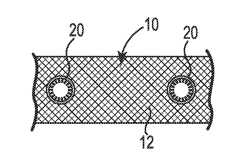
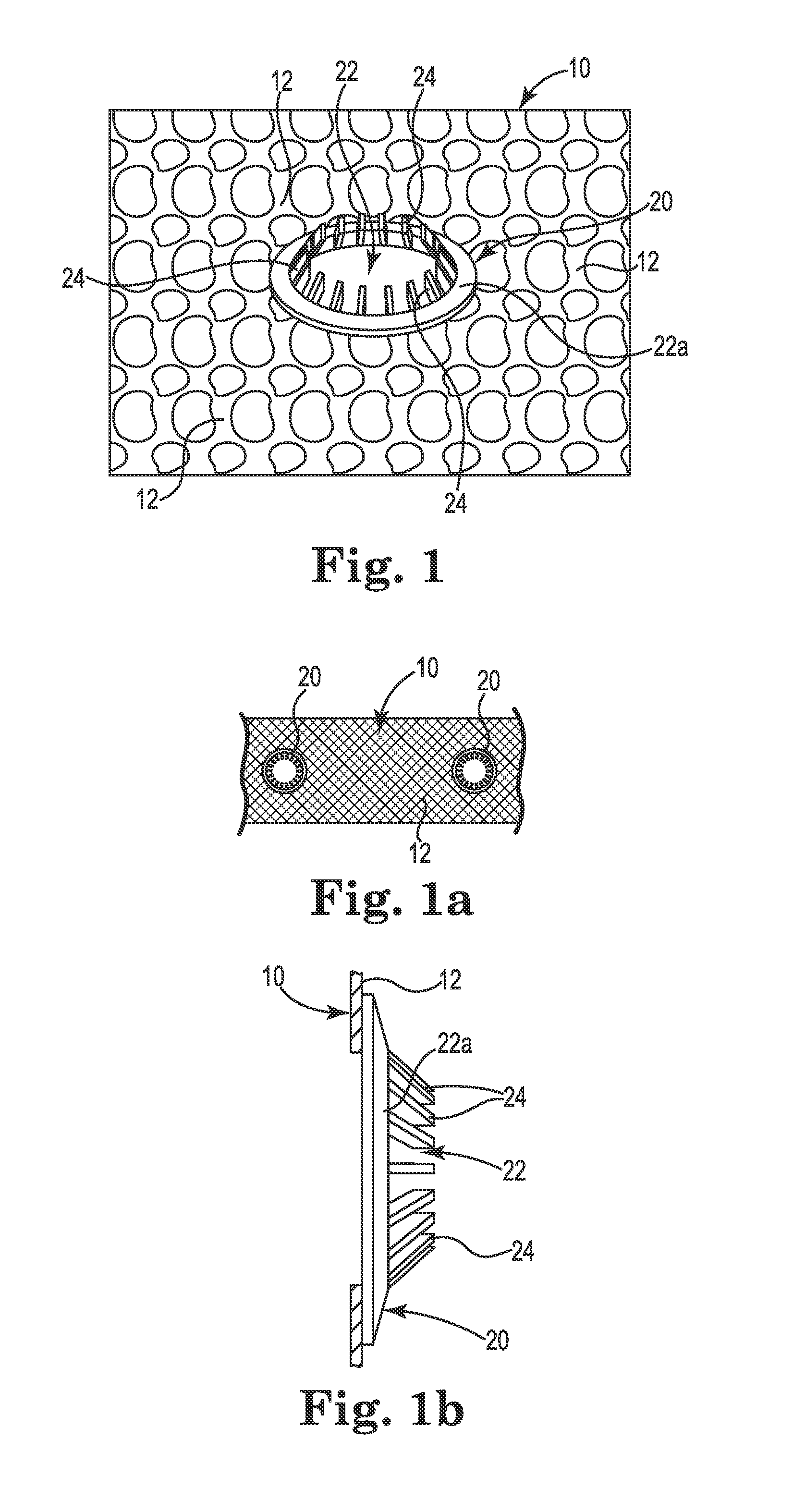
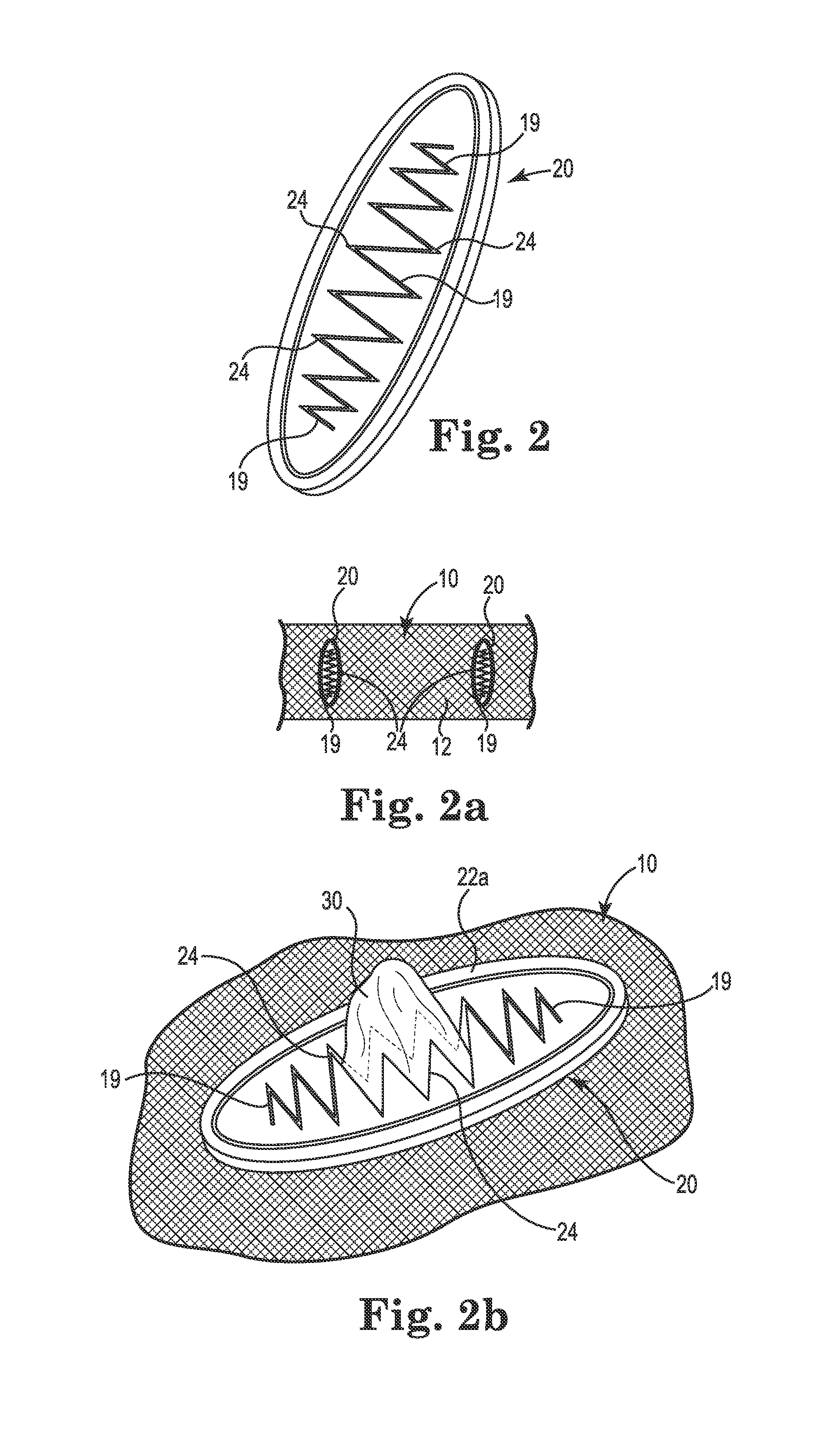
[0018]Referring generally to FIGS. 1-2b, various embodiments of implantable sling or mesh support systems 10 and implant methods are shown. In general, the implant systems 10 can include support portions 12, anchoring portions, and extensions. Various portions of the implant systems 10 can be constructed of polymer materials, including mesh constructs including a plurality of filaments or members to define a lattice form. Certain embodiments can be constructed of or from a film or sheet material of polypropylene, polyethylene, fluoropolymers or like compatible materials, e.g., into a molded generally planar structure or from a thin generally planar film or sheet material. Examples of acceptable polymer materials available in constructing or forming the implant systems and its components can include polypropylene, polyethylene, fluoropolymers or like biocompatible materials.

[0019]The various implants 10 or systems, features and methods detailed herein are envisioned for use with many...


PUM


Abstract

Various embodiments of a mesh or implant system are provided. In certain embodiments, a tissue fixation device is included. The tissue fixation device can include an eyelet structure having an opening and one or more teeth adapted to grasp tissue to secure the implant in place.

Description

PRIORITY AND RELATED APPLICATION[0001]This Application claims priority to and the benefit of U.S. Provisional Patent Application No. 61 / 748,826, filed Jan. 4, 2013, which is hereby incorporated fully herein by reference.FIELD OF THE INVENTION[0002]The present invention relates generally to surgical methods and apparatus and, more specifically, to implantable mesh, sling or implant devices for use in treating incontinence or other pelvic disorders.BACKGROUND OF THE INVENTION[0003]Pelvic health for men and women is a medical area of increasing importance, at least in part due to an aging population. Examples of common pelvic ailments include incontinence (e.g., fecal and urinary), pelvic tissue prolapse (e.g., female vaginal prolapse), and conditions of the pelvic floor.[0004]Urinary incontinence can further be classified as including different types, such as stress urinary incontinence (SUI), urge urinary incontinence, mixed urinary incontinence, among others. Other pelvic floor diso...

Claims


Application Information

Patent Timeline
Patent Type & Authority Applications(United States)
IPC IPC(8): A61F2/00
CPCA61F2/0063A61F2/0045
Inventor SJOQUIST, SCOTT L.ARCAND, BENJAMIN Y.
Owner BOSTON SCI SCIMED INC
Why Eureka
Try Eureka