Medical Device Introduction Systems and Methods

a technology for medical devices and introduction systems, applied in the field of medical device introduction systems and methods, can solve the problems of limiting the ability of physicians to perform as well as capable, limiting the ability of physicians to view a target site, limiting the ability of construction of medical devices, and limiting the ability of physicians to perform additional procedures. to achieve the effect of improving steering

Inactive Publication Date: 2014-07-17
UVISION INC
View PDF3 Cites 66 Cited by
  • Summary
  • Abstract
  • Description
  • Claims
  • Application Information

AI Technical Summary

Benefits of technology

[0011]In some embodiments, one or more of the introducer tube, the working channel tube, and the endoscopic cannula can include a proximal portion having a first durormeter and a distal portion having a second durometer. The second durometer is lower than the first durometer so as to allow deflection of the distal portion of the respective tube or cannula for controllable access to a target area in the interior body region. In some embodiments further durometer(s) may be utilized to enhance steering.
[0015]The present invention can include embodiments of a method. For example, a medical introducer comprising a handle and an elongate introducer tube extending therefrom and having a plurality of lumens extending longitudinally therein can be inserted into an interior body region of a patient. A separate imaging device can be inserted through the handle and in a predetermined one of the plurality of lumens. The imaging device can be positioned in a selected position within the interior body region. Then, an image can be produced from within the interior body region. A separate working channel device and position, controller can be removably connected to the medical introducer. The working channel device can include an elongate working channel tube having at least one lumen extending the length thereof defining a working channel. The position controller for controlling the position of the working channel tube can be positioned in the working channel in another predetermined one of the plurality of lumens. In such embodiments, one of the group of the medical introducer, the imaging device, and the working channel device may be moved independently of the others of the group.
[0016]In some embodiments of a method, the medical introducer handle can comprise an oval-shaped ring of material having an open interior. The method can further include connecting a distal end of the handle to the introducer tube. In some embodiments of a method, the medical introducer can include a modular manifold integrally formed on a proximal end of the introducer tube and have a corresponding plurality of lumens aligned with the plurality of lumens in the introducer tube. In such an embodiment, the manifold can be removably connected to the introducer handle. The manifold and introducer tube may be interchanged in the handle with other manifolds and introducer tubes.

Problems solved by technology

As a result, operating within small spaces with a plurality of medical devices, such as scopes, dilating and cutting instruments, fluids, catheters, implants, and the like, can become difficult to manage.
When performing a procedure with a plurality of medical devices, positioning, controlling, manipulating, and handling the various medical devices during the procedure can limit a physician's ability to perform as well as capable.
That is, the design and construction of a medical device can limit a physician's ability to view a target site, maneuver within a space, transition between procedures, and / or perform additional procedures.
Managing the use of multiple devices in a procedure can pose even greater difficulty to a single physician who desires to perform a procedure, often without assistance or with limited assistance, in an office or outpatient setting so as to avoid the time and expense of hospital utilization for such procedures.
The optical capabilities can be limited due to various factors, including, for example, the anatomical structures about which the scopes are maneuvered, and the movement and / or control together of both the imaging device and a delivery device and the resulting loss in orientation in an interior body region.
For example, optical capabilities with conventional endoscopes typically used in hysteroscopy procedures are often limited in such ways, making it difficult for the physician to know whether what is being viewed is up or down.
Such conventional endoscopes and associated delivery devices are often complex and require extended learning to operate effectively.
In addition, many conventional endoscopes and delivery devices are reusable and can be very expensive to purchase and to re-sterilize after each use.
However, some surgical devices contain very small and / or narrow working channels or lumens for performing intricate medical procedures.
These small and / or narrow working channels can be difficult to clean and sterilize.
If not effectively eliminated, these materials may be transferred to, and potentially cause harmful infections to, other patients or medical personnel through subsequent use of the devices.
In addition to the problems of potential disease transmission and lack of disposability, conventional reusable medical introducer, endoscopes, and the like are subjected to repeated use over prolonged periods.
Further, these devices are regularly subjected to sterilization with heat or chemicals.
To accomplish these objectives, conventional reusable devices are often made of stainless steel or other durable materials that are costly.
In addition, despite being designed for repeated use, such conventional intricate reusable devices, in particular, such devices that incorporate visualization components, often require regular replacement, further adding to the cost of such devices.

Method used


Image

  • Medical Device Introduction Systems and Methods
  • Medical Device Introduction Systems and Methods
  • Medical Device Introduction Systems and Methods

Examples


Embodiment Construction

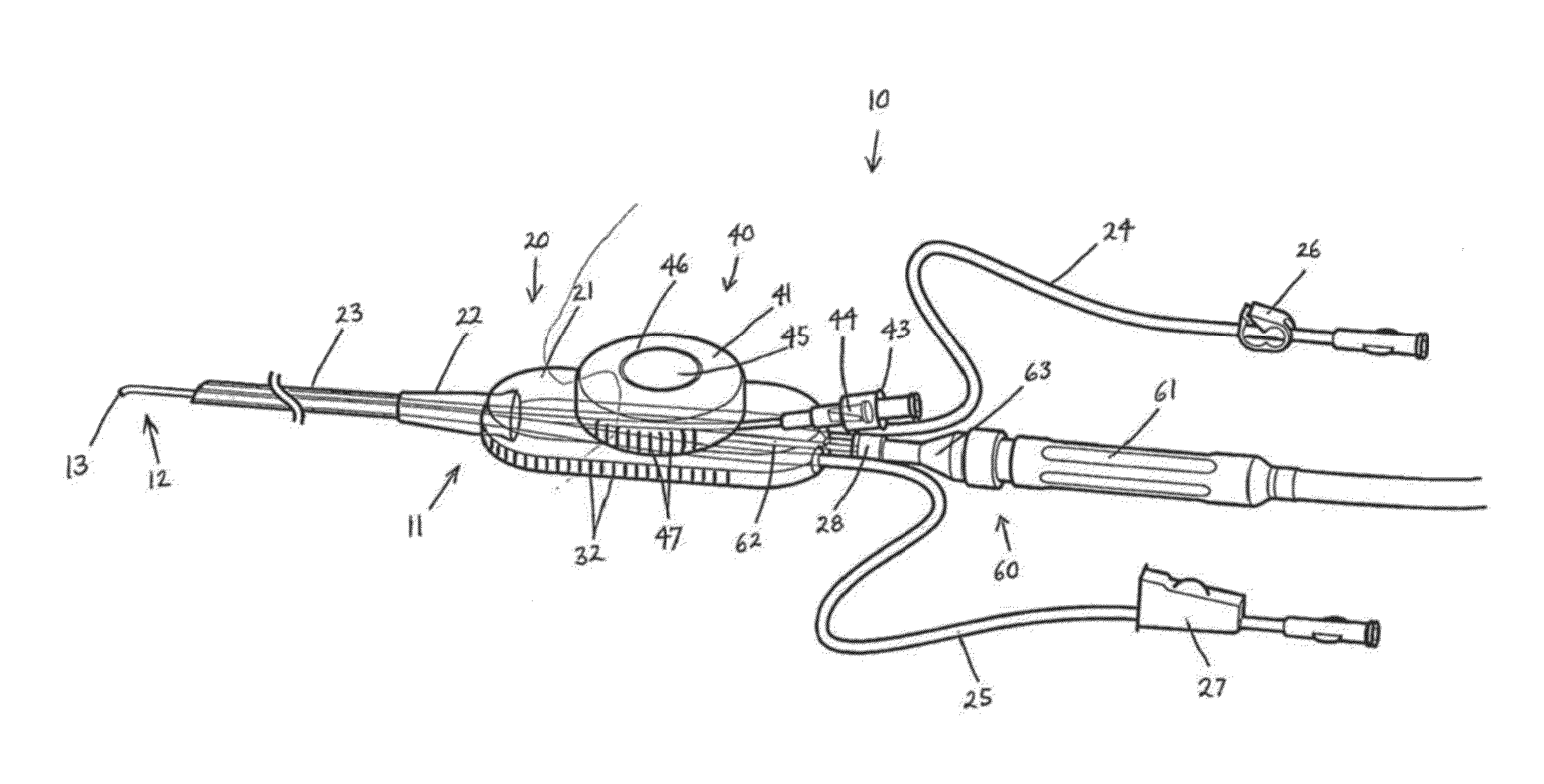
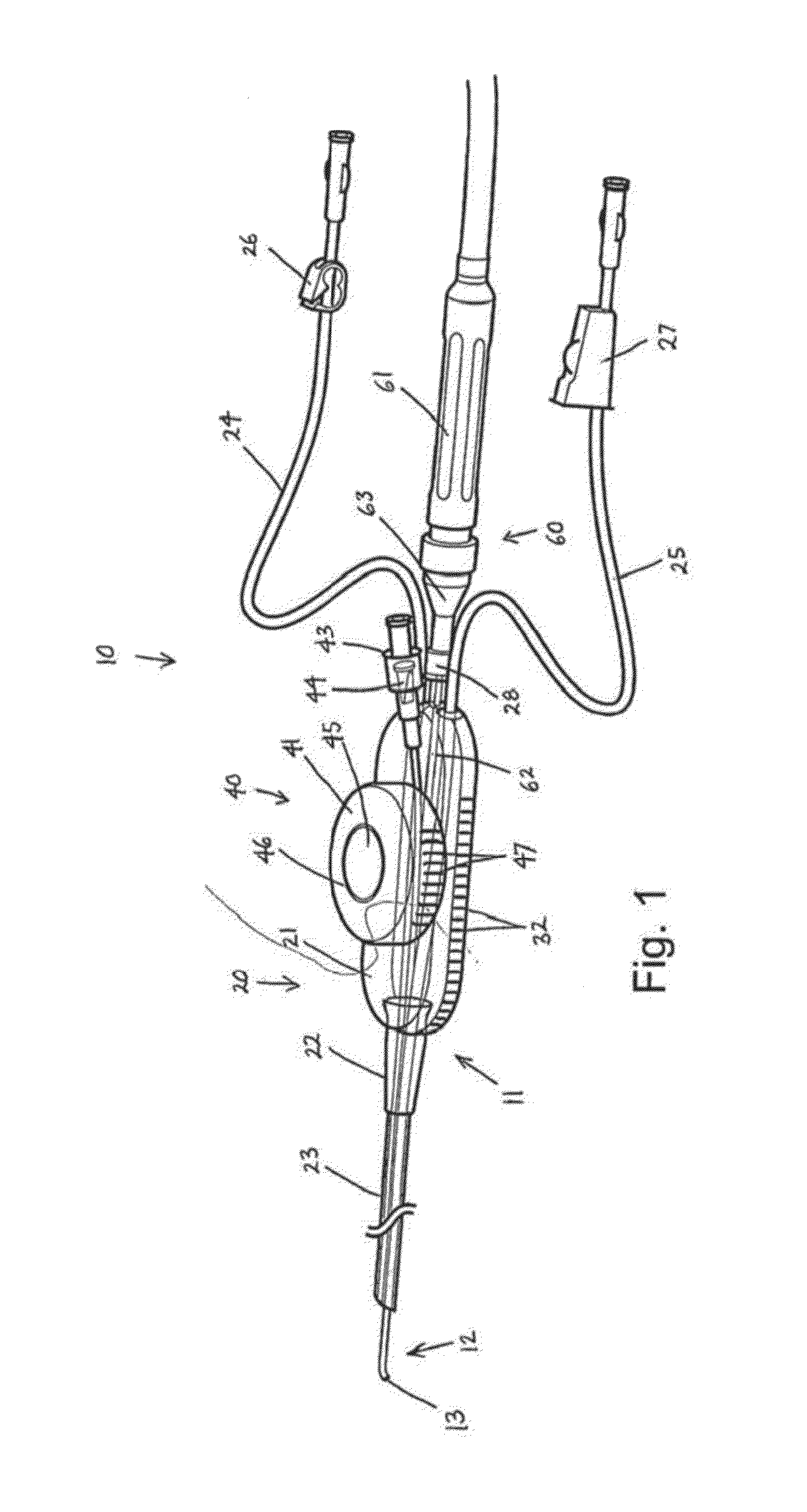
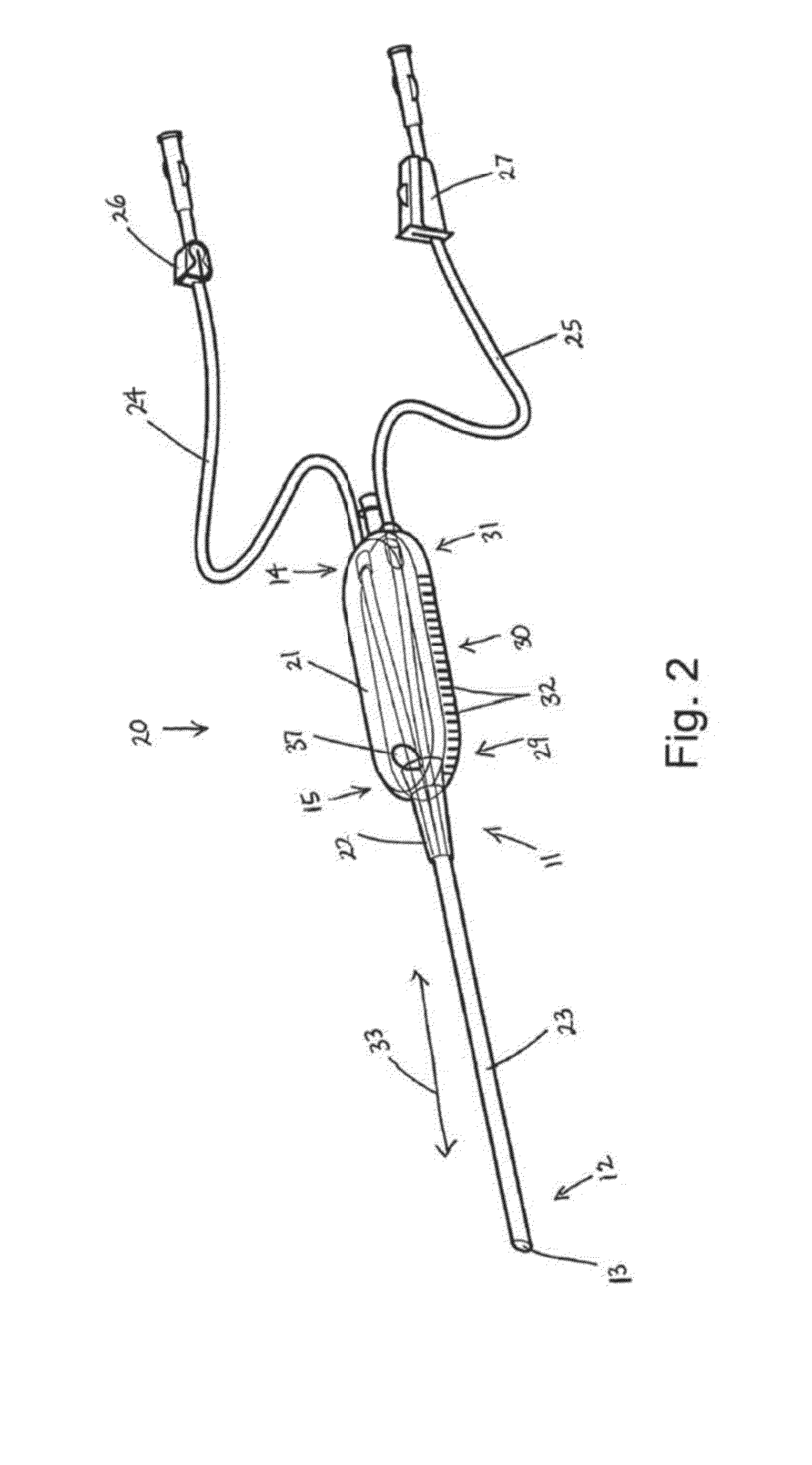
[0051]Some embodiments of the present invention can provide a medical device introduction system and / or method. FIGS. 1-18 show various aspects of such embodiments. For example, an illustrative embodiment of a medical device introduction system and / or method can include a medical introducer, a separate imaging device, and / or a separate working channel device. In such an embodiment, each of the medical introducer, the imaging device, and the working channel device can be movable independent of the other.

[0052]Minimally invasive surgical procedures have been developed that can be used in many diagnostic and / or therapeutic medical procedures. Such minimally invasive procedures can reduce pain, post-operative recovery time, and the destruction of healthy tissue. In minimally invasive surgery, the site of pathology can be accessed through portals rather than through a significant incision, thus preserving the integrity of intervening tissues. These minimally invasive techniques also ofte...


PUM


Abstract

A medical device introduction system and method can include a medical introducer, a separate imaging device, and / or a separate working channel device, each of which may be movable independent of the other. The medical introducer can include a handle and an elongate introducer tube extending from the handle and having a plurality of lumens, and may be inserted into an interior body region of a patient. The separate imaging device may be inserted through the handle and positioned in one of lumens. The separate working channel device can include an elongate working channel tube and a position controller. The working channel tube can include at least one lumen defining a working channel. The position controller can be configured to control positioning of the working channel tube. The working channel device may be removably connected to the handle and positioned in another lumen. Associated with the medical device may be a slider and a rail that stabilizes a medical device and prevents it from turning allowing medical personnel that previously had to hold the medical device to be free to be able to perform other tasks. The medical device may also possess a bent working channel device that provides easier access to orifices or other body parts that are in an orientation that is not straight (i.e., in an orientation that does not require a bend in the working channel device). The medical device may also have sheaths associated with it that causes a bend in the working channel device that also allows easier access to orifices or other body parts that are not straight

Description

[0001]This application claims priority under 35 USC 119(e) to U.S. Provisional Application No. 61 / 753,413 filed Jan. 16, 2013, the contents of which are incorporated by reference in its entirety.FIELD OF THE INVENTION[0002]The present invention relates to medical device introduction systems and methods. Embodiments of the present invention may be useful for separately introducing and independently controlling multiple cooperating medical devices in interior body regions.BACKGROUND OF THE INVENTION[0003]In recent years, medical procedures have advanced to stages where less and less invasive, or minimally invasive, surgeries, diagnostic procedures, exploratory procedures, or other medical procedures have been desired and demanded by patients, physicians, and payers. To accomplish these desires and demands, various medical devices and instrumentation have been developed, such as cannulas or micro-cannulas, various catheter devices, micro-surgical instrumentation and implants, medical i...

Claims


Application Information

Patent Timeline
Patent Type & Authority Applications(United States)
IPC IPC(8): A61B1/018A61B1/015A61B1/00A61B17/00A61B1/04
CPCA61B1/018A61B1/00066A61B17/00234A61B1/015A61B1/04A61B1/00154A61B17/42A61B2017/003A61B2017/4216A61B2017/4233A61B2218/002A61B2218/007A61B1/0052A61B2090/306A61B1/00165A61B1/002A61B1/0661A61B1/00016A61B1/0011A61B1/05A61B1/0669A61B1/0684A61B1/07H04N23/555H04N23/45A61B1/005A61B1/00018A61B1/00029A61B1/00045A61B1/00121A61B1/0676A61B1/051A61M25/003
Inventor SNOKE, PHILLIP JACKALLRED, PHILIP MORRISON
Owner UVISION INC
Why Eureka
Try Eureka