Ultrasound diagnostic imaging apparatus

a diagnostic imaging and ultrasonic technology, applied in diagnostics, medical science, applications, etc., can solve the problems of increasing the size of the apparatus and the inability to intuitively operate, and achieve the effect of less space consumption

Active Publication Date: 2014-08-07
KONICA MINOLTA INC
View PDF3 Cites 28 Cited by
  • Summary
  • Abstract
  • Description
  • Claims
  • Application Information

AI Technical Summary

Benefits of technology

[0008]The present invention has been made in consideration of the above problems, and it is one of main objects to provide an ultr...

Problems solved by technology

However, the following problems occur when the ultrasound diagnostic imaging apparatus which is able to execute the dual screen display mode where the display region is divided between an upper display region and a lower display region, and the dual screen display mode where the display region is divided between a left display region and a right display region is configured to be operated by only three keys (for example, mode switching key: one, dis...

Method used


Image

  • Ultrasound diagnostic imaging apparatus
  • Ultrasound diagnostic imaging apparatus
  • Ultrasound diagnostic imaging apparatus

Examples


first embodiment

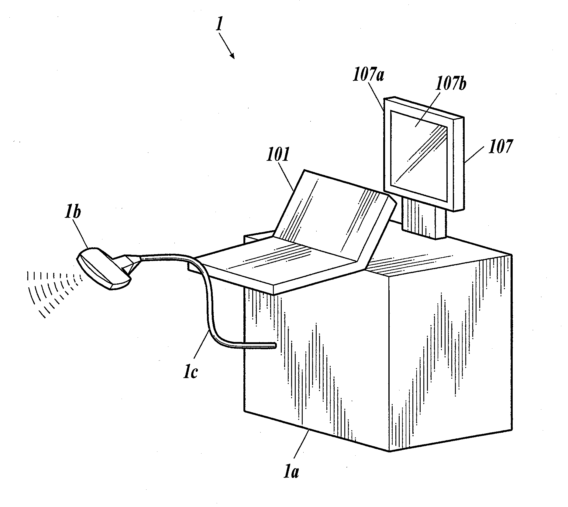
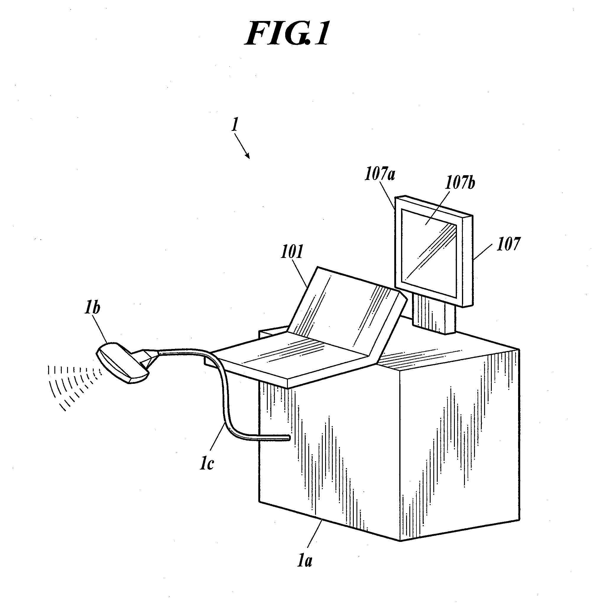
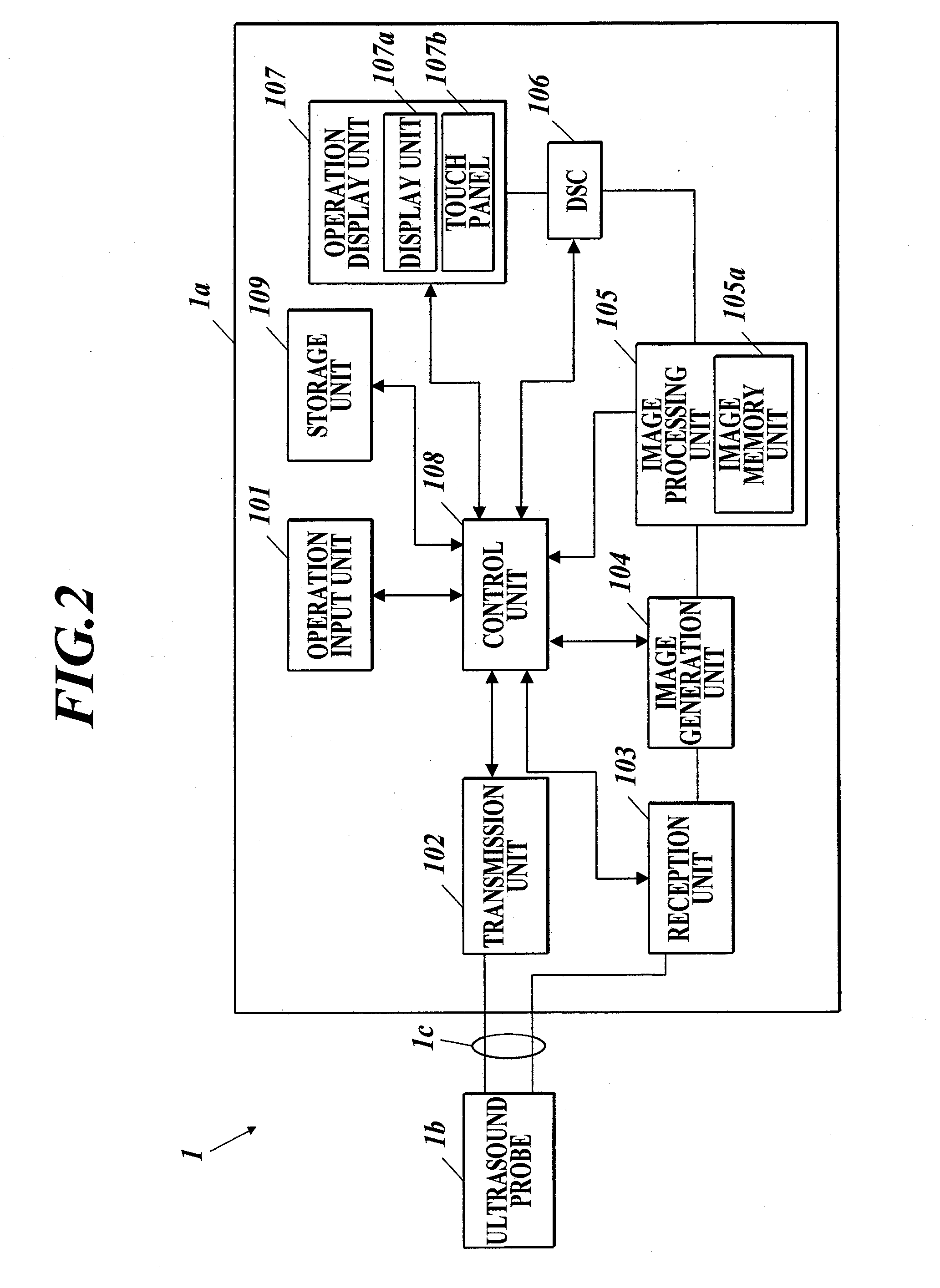
[0067]As shown in FIG. 1 and FIG. 2, the ultrasound diagnostic imaging apparatus 1 of the first embodiment includes an ultrasound diagnostic imaging apparatus main body 1a and an ultrasound probe 1b. The ultrasound probe 1b transmits ultrasound (transmitting ultrasound) to a test subject such as a live body which is not shown, and receives a reflecting sound (reflecting ultrasound: echo) of the ultrasound reflecting on the test subject. The ultrasound diagnostic imaging apparatus main body 1a is connected to the ultrasound probe 1b through a cable 1c. The ultrasound diagnostic imaging apparatus main body 1a transmits a driving signal which is an electric signal to the ultrasound probe 1b so that the ultrasound probe 1b transmits the transmitting ultrasound to the test subject. Then, the ultrasound diagnostic imaging apparatus main body 1a images an inner state of the test subject as an ultrasound image based on a receiving signal which is an electrical signal generated by the ultras...

second embodiment

[0118]Next, the second embodiment is described. In the second embodiment, when the display format advances to the dual screen display mode, the active display region is switched by directly touching the display region. With such configuration, according to the second embodiment, it is possible to switch the active display region more intuitively.

[0119]The specific configuration of the ultrasound diagnostic imaging apparatus 1 of the second embodiment is similar to the ultrasound diagnostic imaging apparatus 1 of the first embodiment. Therefore, the same reference numerals are applied and the description is omitted.

[0120]For example, in the second embodiment, when the scanning operation starts as described in the first embodiment, if the dual display mode advancing button B4 is touched, the display format advances from the single display mode to the dual screen display mode or the quadruple screen display mode.

[0121]For example, in a case where the single display mode advances to the...


PUM


Abstract

Disclosed is an ultrasound diagnostic imaging apparatus which displays an ultrasound image on a display screen of a display unit based on ultrasound image data. The ultrasound diagnostic imaging apparatus includes a touch panel and a control unit. The control unit is able to execute a vertical display mode to display two ultrasound images aligned vertically on the display screen and a horizontal display mode to display two ultrasound images aligned horizontally on the display screen. In each display mode, the control unit sets operation reception regions aligned vertically or horizontally on the touch panel. When the control unit detects an operation of touching one of the operation reception regions, the control unit sets an ultrasound image displayed in the touched side to a selected state.

Description

BACKGROUND[0001]1. Field of the Invention[0002]The present invention relates to an ultrasound diagnostic imaging apparatus.[0003]2. Description of Related Art[0004]Conventionally, there is known an ultrasound diagnostic imaging apparatus which is able to execute two modes where the modes are a single display mode in which one ultrasound image is displayed in an ultrasound image display region on a display screen of a display unit and a dual screen display mode in which the ultrasound image display region is divided in half vertically or horizontally, and two ultrasound images are displayed, one in each display region (for example, the above is described in Japanese Patent Application Laid-Open Publication No. 2001-276062).[0005]When image adjustment, measurement, etc. are performed on the ultrasound image displayed in each display region in the dual screen display mode, it is useful to be able to perform the above operation for each display region. Therefore, it is necessary to swit...

Claims


Application Information

Patent Timeline
IPC IPC(8): A61B8/00
CPCA61B8/463A61B8/44A61B8/465A61B8/468A61B8/469A61B8/54
Inventor OTA, KAZUSHI
Owner KONICA MINOLTA INC
Why Eureka
Try Eureka