Method for quantifying 5-hydroxymethylcytosine

a technology of cytosine and cytosine base, which is applied in the field of methods for detecting and quantifying cytosine bases, can solve the problems of cumbersome techniques and inability to accurately quantify the amount of cytosine bases

Inactive Publication Date: 2014-09-18
PROMEGA CORP
View PDF0 Cites 19 Cited by
  • Summary
  • Abstract
  • Description
  • Claims
  • Application Information

AI Technical Summary

Problems solved by technology

Unfortunately, these techniques are often cumbersome as they rely on the use of antibodies in multi-step ELISA techniques, on the use of multiple washes and / or the use of radioactivity.

Method used


Image

  • Method for quantifying 5-hydroxymethylcytosine
  • Method for quantifying 5-hydroxymethylcytosine
  • Method for quantifying 5-hydroxymethylcytosine

Examples


example 1

Materials Relating to Examples 2-5

[0102]All DNA samples used in the below examples were purchased from Active Motif or ZYMO Research. T4 β-Glucosyltransferase was either purchased from ZYMO Research or recombinantly produced at Promega Corporation. The UDP Detection Reagent contains Promega Glo buffer with UDP converting enzyme (CMK) and luciferase / luciferin component. TET1 enzyme was purchased from Active Motif

example 2

Converting 5-hmC to Luminescence

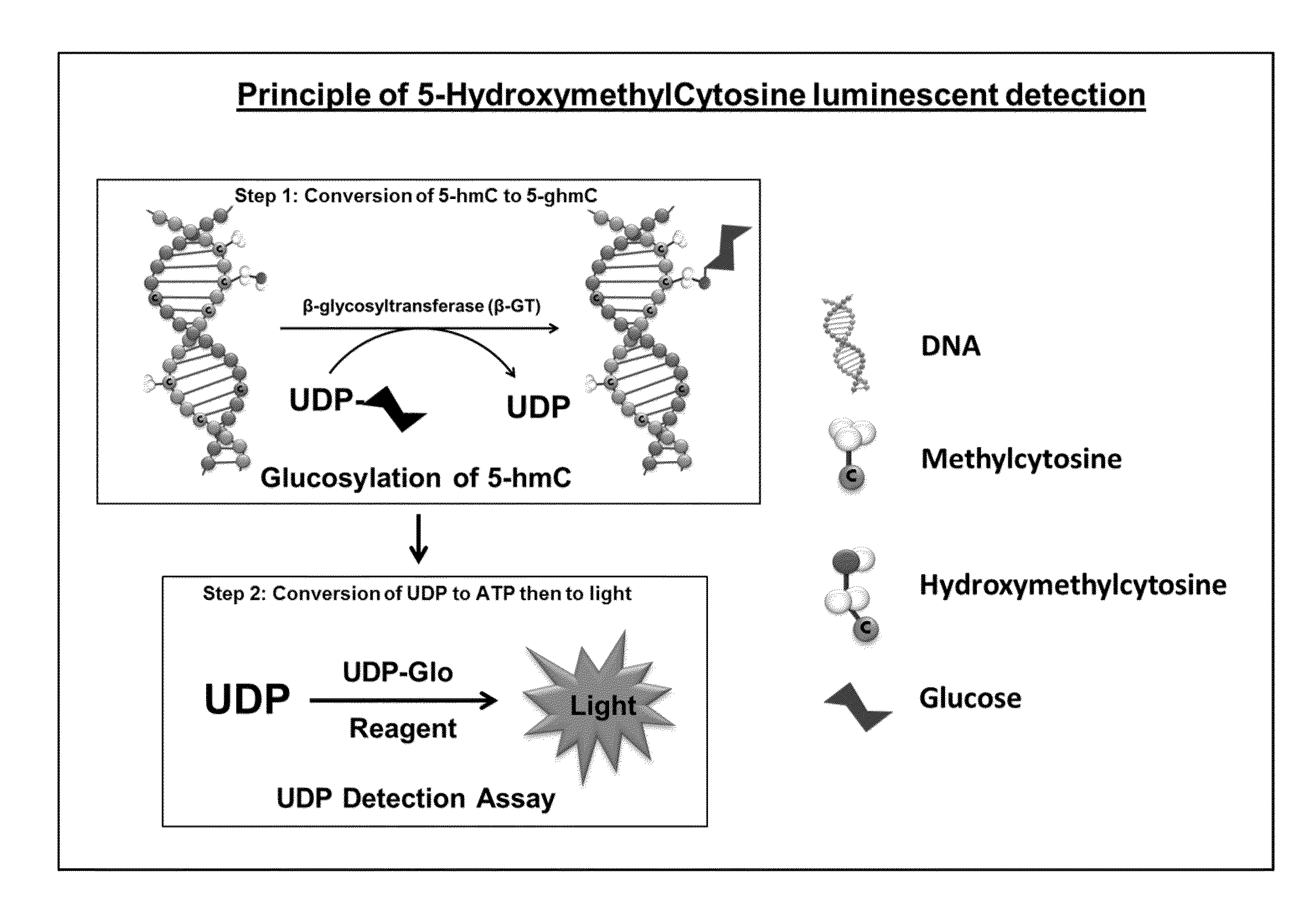
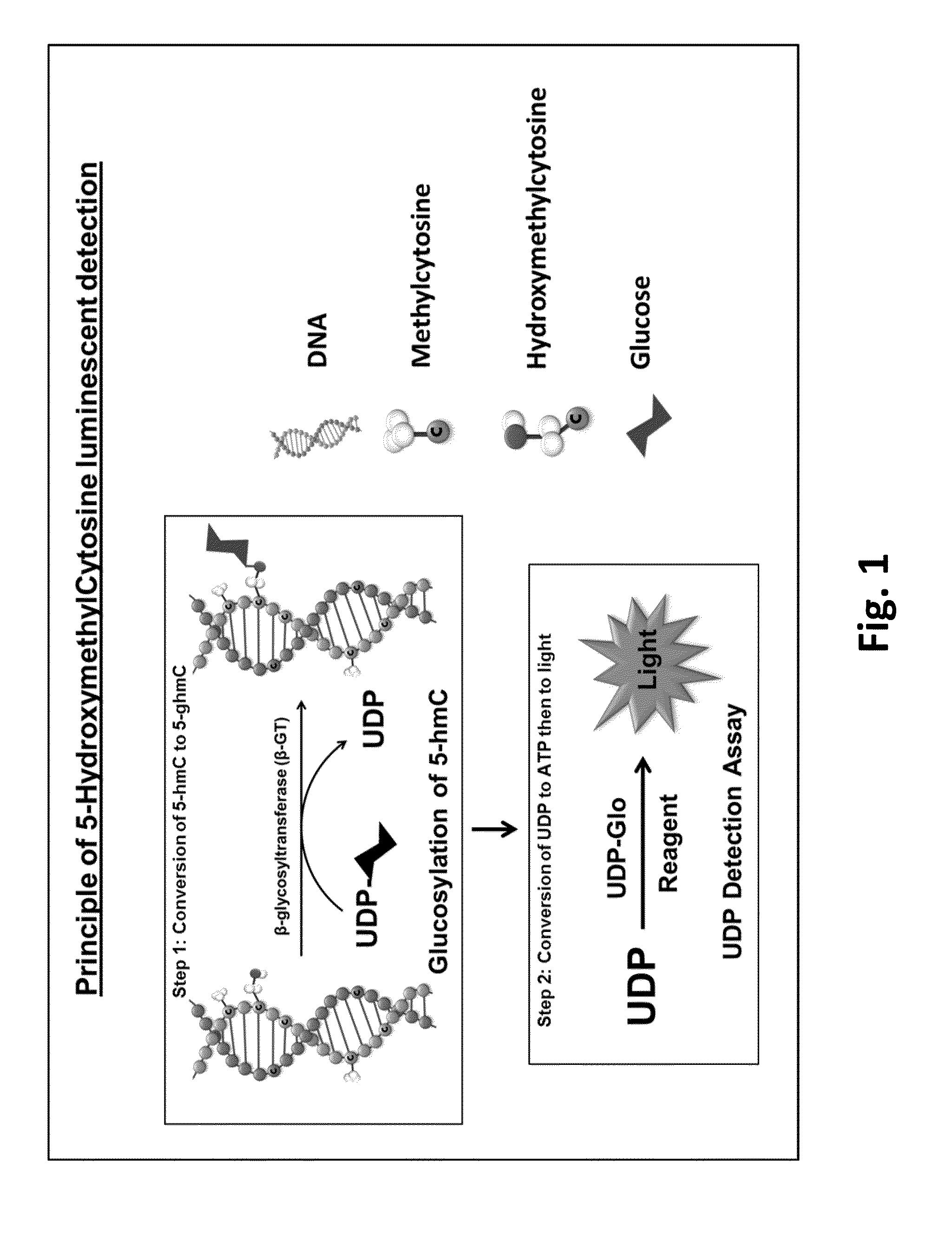
[0103]DNA containing a known amount of cytosines was used in this experiment. DNA with all cytosines unmodified, methylated or hydroxymethylated were serially diluted in multi-well plates starting from 100 ng DNA in 25 μl of total reaction volume. Reactions were performed in GT Buffer (10 mM Tris pH 7.5, 10 mM NaCl, 10 mM MgCl2 and 1 mM DTT) containing 0.125 U β-GT and 50 mM UDP-glucose donor substrate. The glucosyltransferase reaction was performed at 37° C. for 1 hour. To detect the UDP produced, 25 μl of UDP Detection Reagent was added to convert UDP to ATP and then ATP to light. After an hour, the luminescence generated was measured on a luminometer.

[0104]As shown in FIG. 3, the 5-hmC detection signal was linear and proportional to the amount of 5-hmC in the sample. Only the 5-hmC containing DNA was able to be glucosylated by β-GT and hence generated UDP. The unmethylated and methylated cytosines could not be glucosylated by the glycosyltransferas...

example 3

Luminescence is Proportional to UDP and 5-hmC

[0106]UDP was titrated in 25 μl GT buffer to create a standard curve. 25 μl of UDP detection reagent was added, and after 1 hour at room temperature, luminescence was measured on a luminometer. Luminescence generated from 5-hmC conversion as described in Example 1 was converted to concentration of UDP formed based on the UDP standard curve.

[0107]FIG. 5 shows that luminescence is proportional to the UDP produced, and, by using the UDP standard curve, there is a positive correlation between the amount of 5-hmC DNA and the UDP produced by β-GT.


PUM

PropertyMeasurementUnit
molecular weightaaaaaaaaaa
molecular weightsaaaaaaaaaa

Abstract

Provided herein are methods for detecting and quantifying 5-hydroxymethylated cytosine bases in a DNA molecule.

Description

CROSS REFERENCE TO RELATED APPLICATIONS[0001]This application claims priority to U.S. Provisional Application No. 61 / 793,936, filed Mar. 15, 2013, which is incorporated herein by reference in its entiretyFIELD OF THE INVENTION[0002]The present invention relates to the development of methods for detecting and quantifying 5-hydroxymethylated cytosine bases in a DNA molecule.BACKGROUND[0003]Methylation of DNA is catalyzed by DNA methyltransferases and occurs at the 5-carbon position of cytosine residues to form 5-methylcytosines (5-mC). 5-mC constitutes approximately 2-8% of the total cytosines in human genomic DNA and influences a broad range of biological functions. These functions include gene expression, maintenance of genomic integrity, parental imprinting, X-chromosome inactivation, regulation of development, aging and cancer.[0004]Ten-Eleven Translocation proteins (TETs) drive the oxidation of 5-mC. The resultant 5-hydroxymethylcytosines (5-hmC) may play critical roles in the pa...

Claims


Application Information

Patent Timeline
Patent Type & Authority Applications(United States)
IPC IPC(8): C12Q1/66
CPCC12Q1/66C12Q1/6827C12Q1/48C12Q1/485G01N2333/90245C12Y204/01028C12Y207/04022C12Q2563/103
Inventor ZEGZOUTI, HICHAMENGEL, LAURIEGOUELI, SAID A.
Owner PROMEGA CORP
Why Eureka
Try Eureka