

Magnetic resonance imaging method for imaging components with short transverse relaxation times (T2) in a human or an animal heart
a technology of magnetic resonance imaging, which is applied in the field of magnetic resonance imaging (mri) method for imaging components with short transverse relaxation time (t2) in a human, can solve the problems of hardly being used in clinical routine, prone to misinterpretation of obtained magnetic resonance images, and radial acquisition scheme which not only requires an extended sampling time,
- Summary
- Abstract
- Description
- Claims
- Application Information
AI Technical Summary
Benefits of technology
Problems solved by technology
Method used
Image

Examples
Embodiment Construction
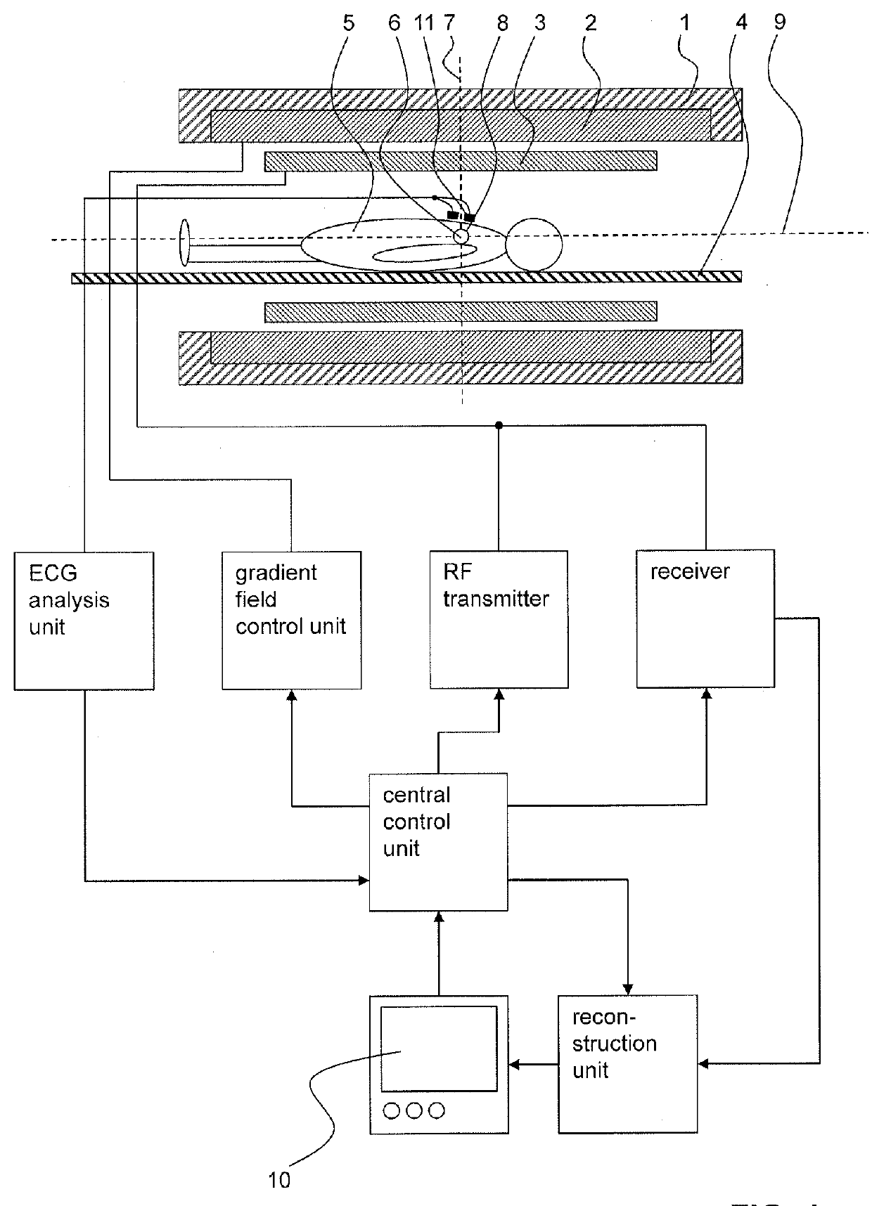
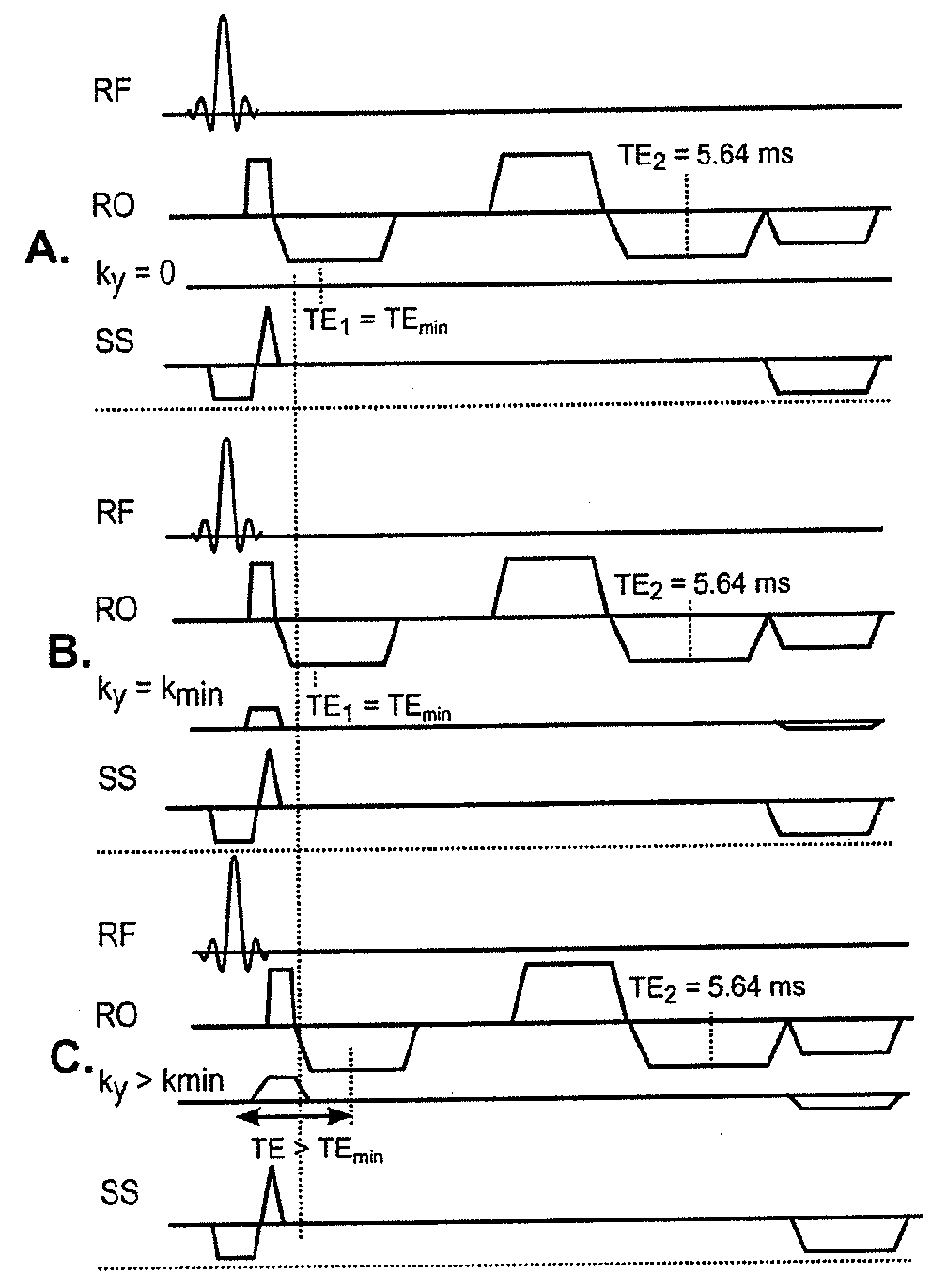
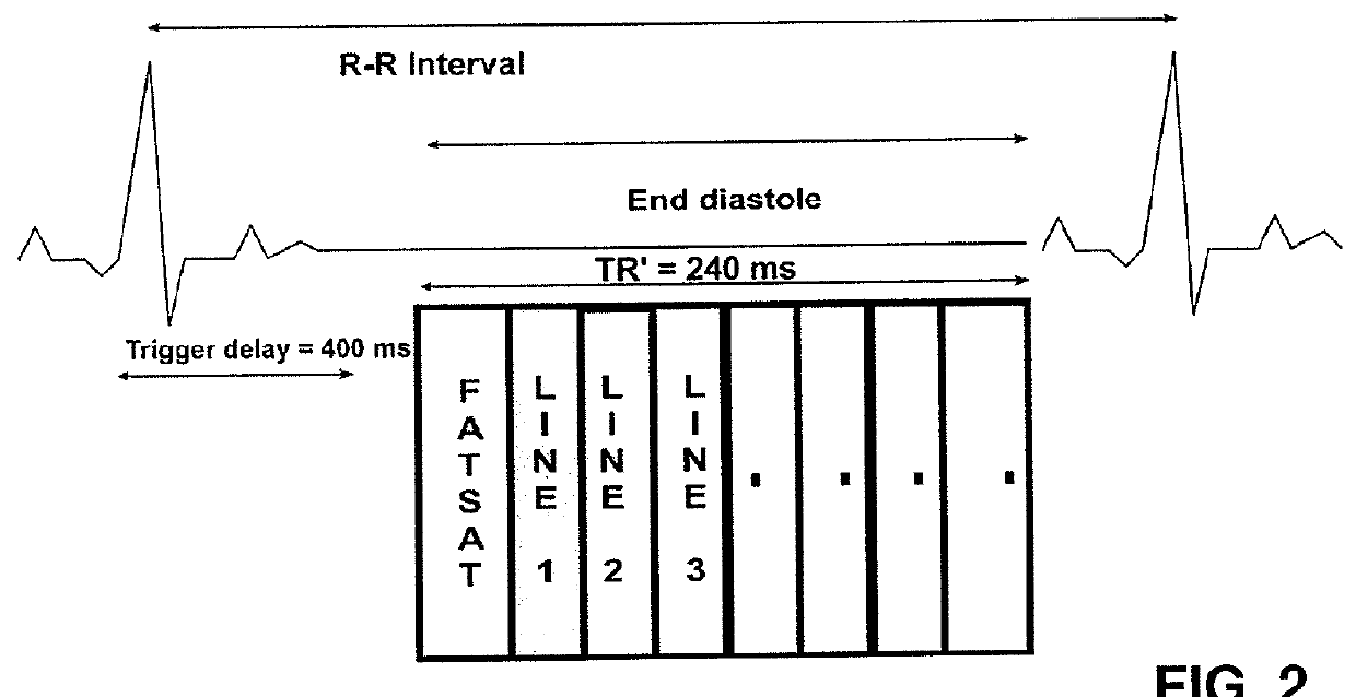
[0070]In FIG. 1, an exemplary MRI system is shown which serves to carry out the inventive method for imaging components with short transverse relaxation times (T2) in a human or an animal heart. Preferred embodiments of the inventive method are schematically illustrated in FIGS. 2-4.
[0071]The MRI system comprises a main magnet 1 for producing a main magnetic field B0. The main magnet 1 usually has the essential shape of a hollow cylinder with a horizontal bore. Inside the bore of the main magnet 1 a magnetic field is present, which is essentially uniform at least in the region of the isocenter 6 of the main magnet 1. The main magnet 1 serves to at least partly align the nuclear spins of a sample 5 arranged in the bore. Of course, the magnet 1 does not necessarily be cylinder-shaped, but could for example also be C-shaped.
[0072]A patient 5 is arranged in such a way on a moving table 4 in the bore of the main magnet 1, that the heart of the patient 5, of which components with short tr...
PUM
 Abstract
Description
Claims
Application Information