

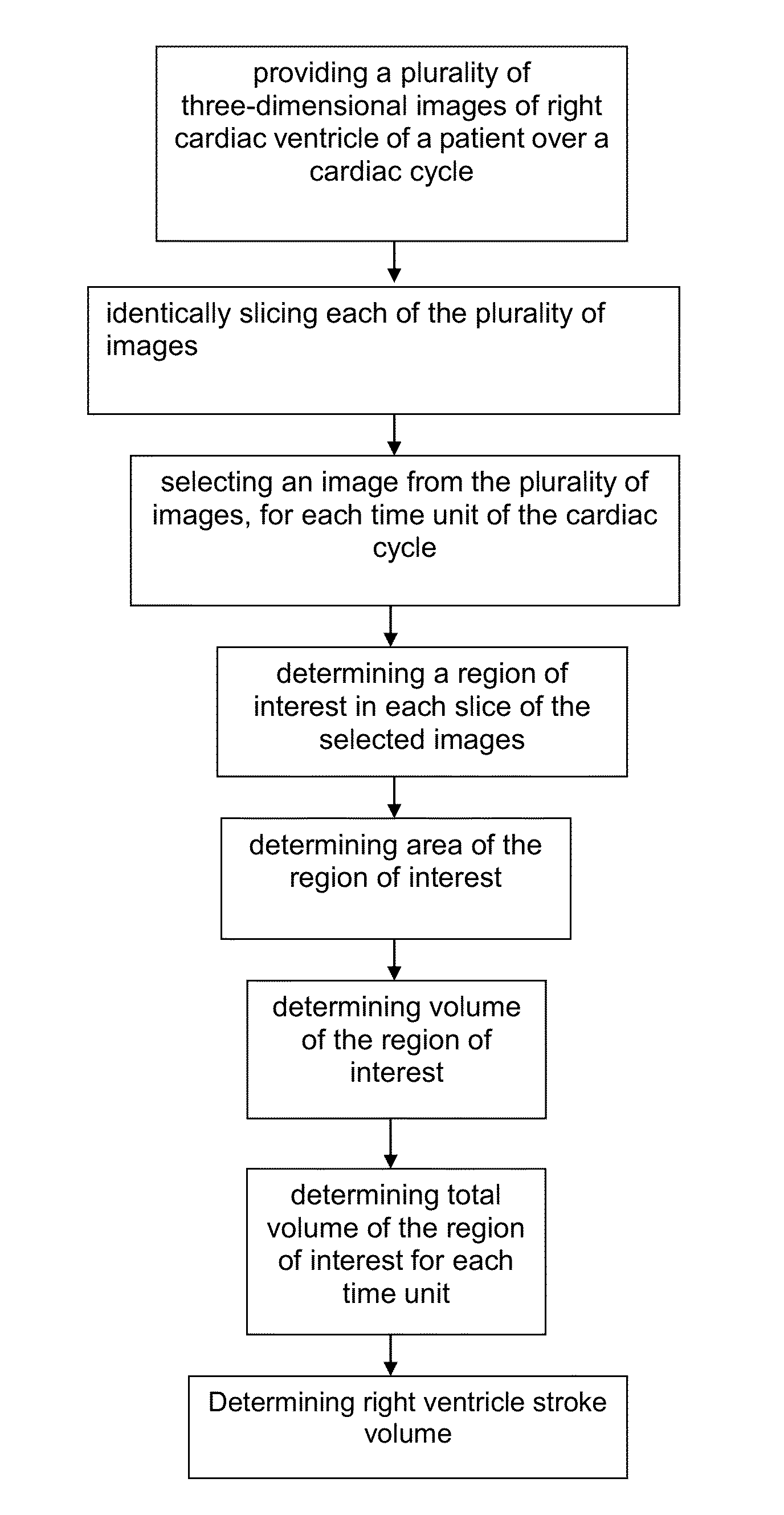
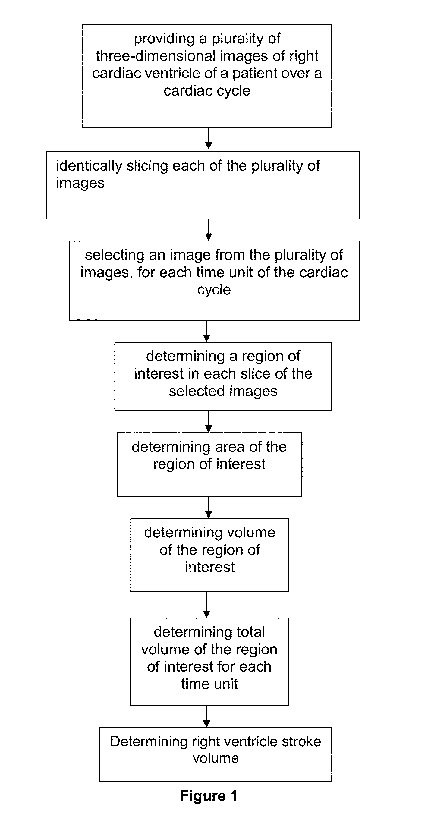
Method for Determining Right Ventricle Stroke Volume
a right ventricle and volume measurement technology, applied in the field of measuring the volume of the right ventricle, can solve the problems of discomfort and pain of patients, difficult and inaccurate right ventricle modelling, and the congenital function and anatomy of the ventricle, and achieve the effect of minimal manual manipulation and correction
- Summary
- Abstract
- Description
- Claims
- Application Information
AI Technical Summary
Benefits of technology
Problems solved by technology
Method used
Image

Examples
example
[0041]Below is an example of the method for measuring right ventricle stroke volume.
[0042]In the following example, the input is an image of the right cardiac ventricle of a patient provided by an ultrasound machine. As the parameters given by the ultrasound machine may be inadequate to calculate image resolution, resolution of 3-dimensional transducer of the ultrasound machine is first determined. In a preferred embodiment, the resolution of the input is predetermined using an equation:
resolution=∑i=1pscale(realsizeno.ofpixelsinimage)in(4)[0043]wherein p is iteration number and n is number of iteration.
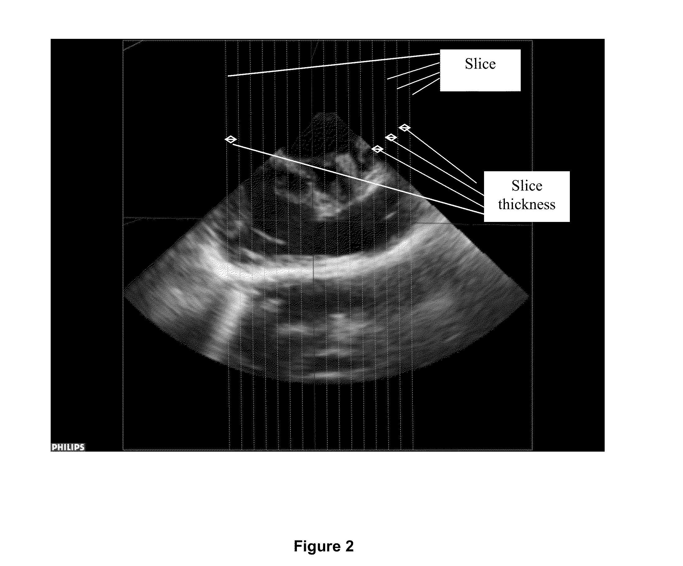
[0044]Determining Resolution of Input
[0045]A test object, for example a piece of meat, is used to determine the resolution of the transducer. The real size of the object is measured. A first ultrasound image is produced using the ultrasound machine.
[0046]The number of pixels in the first image of the object is counted. The object is then moved to change the distance between the objec...
PUM
 Abstract
Description
Claims
Application Information