
Lesion detection and image stabilization using portion of field of view

a technology of image stabilization and lesion detection, applied in the field of endoscopy, can solve the problem of little improvement in the level of achieve the effect of improving the quality of endoscopic procedures

Inactive Publication Date: 2015-03-19
CERNER INNOVATION
View PDF7 Cites 24 Cited by
  • Summary
  • Abstract
  • Description
  • Claims
  • Application Information

AI Technical Summary

Benefits of technology

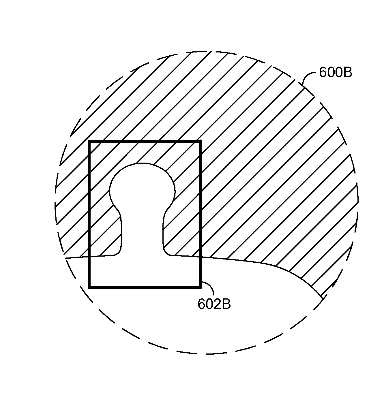
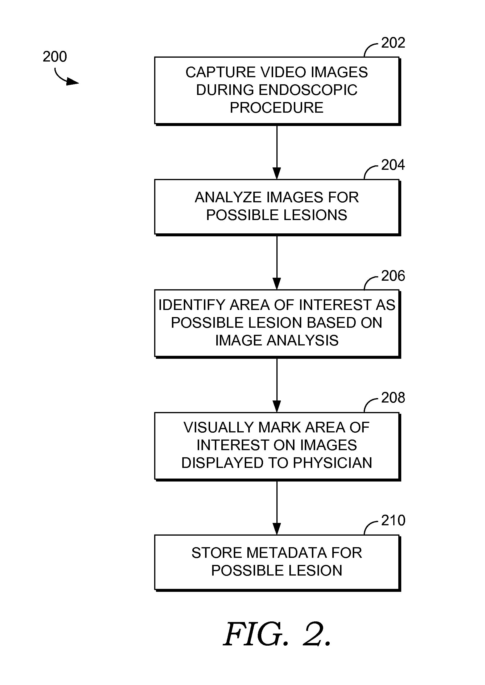
The patent describes techniques to improve endoscopic procedures by using an overall field of view (FOV) to capture images but only displaying a portion of them to an endoscopist during the procedure. The system can identify areas of interest that may contain possible lesions and track their movement within the FOV. This allows for a continuous analysis of the FOV to improve the quality of the endoscopic procedure. The patent highlights the importance of detecting and analyzing areas of interest in order to improve diagnostic accuracy and patient safety.

Problems solved by technology

One of the key challenges in endoscopy is maintaining sufficiently high quality of screening.
However, these debates have done little to actually improve the level of quality of endoscopic procedures.

Method used


Image

  • Lesion detection and image stabilization using portion of field of view
  • Lesion detection and image stabilization using portion of field of view
  • Lesion detection and image stabilization using portion of field of view

Examples


Embodiment Construction

[0023]The subject matter of the present invention is described with specificity herein to meet statutory requirements. However, the description itself is not intended to limit the scope of this patent. Rather, the inventors have contemplated that the claimed subject matter might also be embodied in other ways, to include different steps or combinations of steps similar to the ones described in this document, in conjunction with other present or future technologies. Moreover, although the terms “step” and / or “block” may be used herein to connote different components of methods employed, the terms should not be interpreted as implying any particular order among or between various steps herein disclosed unless and except when the order of individual steps is explicitly described.

[0024]Embodiments of the present invention provide techniques to assist endoscopists in improving the quality of endoscopic procedures. Unless otherwise indicated, these techniques may be employed in real-time ...


PUM


Abstract

Images with an overall field of view (FOV) may be captured during an endoscopic procedure but only a portion of the overall FOV displayed to an endoscopist. The overall FOV may be analyzed to identify an area of interest of a patient's organ based on the presence of a possible lesion during the endoscopic procedure. The endoscopist may be notified of the identified area of interest. After an area of interest is identified, the location of the area of interest may be tracked. Image stabilization may be provided by changing the displayed portion of the overall FOV to maintain the area of interest within the displayed portion as the area of interest moves relative to the endoscope.

Description

CROSS-REFERENCE TO RELATED APPLICATIONS[0001]This application is related by subject matter to the invention disclosed in U.S. Application No. (not yet assigned) (Attorney Docket Number CRNI.189776), filed on even date herewith, entitled “MARKING AND TRACKING AN AREA OF INTEREST DURING ENDOSCOPY,” which is assigned or under obligation of assignment to the same entity as this application, and incorporated in this application by reference.BACKGROUND[0002]One of the key challenges in endoscopy is maintaining sufficiently high quality of screening. Quality for endoscopy is generally defined in part on whether the endoscopist found all lesions that may be of interest during an endoscopic procedure. This may include, for instance, lesions that may be cancerous or pre-cancerous. Various societies debate on different approaches to endoscopic procedures and different ways to evaluate performance of endoscopists. However, these debates have done little to actually improve the level of quality ...

Claims


Application Information

Patent Timeline
IPC IPC(8): G06T7/20A61B1/00
CPCG06T7/20A61B1/00009G06T2207/30096G06T2207/10068G06T2207/10016A61B1/00045A61B5/065A61B2576/00G06T7/0016G06T2207/10021G06T2207/20081G06T2207/20104G06T7/70G16H40/63G16H50/50G16H20/40G16H30/40G16H30/20A61B1/000095
Inventor STAPLES, II, ALAN HARRISRAMSEY, KAREN KAYE
Owner CERNER INNOVATION
Learn More