Method for epicardial pacing or cardiac tissue ablation

a technology of epicardial pacing and cardiac tissue, applied in the field of epicardial pacing or cardiac tissue ablation, can solve the problems of asymmetric or desynchronized left ventricular, affecting the pumping ability of an already weakened left ventricle, and affecting the ability of the left ventricle to pump,

Inactive Publication Date: 2015-07-23
MONTEFIORE MEDICAL CENT INC
View PDF6 Cites 10 Cited by
  • Summary
  • Abstract
  • Description
  • Claims
  • Application Information

AI Technical Summary

Problems solved by technology

This results in an asymmetrical or desynchronized left ventricular contraction that compromises the pumping ability of an already weakened left ventricle.
The major challenge has been placement of the left ventricular lead.
Unfortunately, access to the coronary sinus is sometimes difficult, and many patients do not have a suitable vein branch derived from the coronary sinus.
This procedure is usually undertaken

Method used


Image

  • Method for epicardial pacing or cardiac tissue ablation
  • Method for epicardial pacing or cardiac tissue ablation

Examples


examples

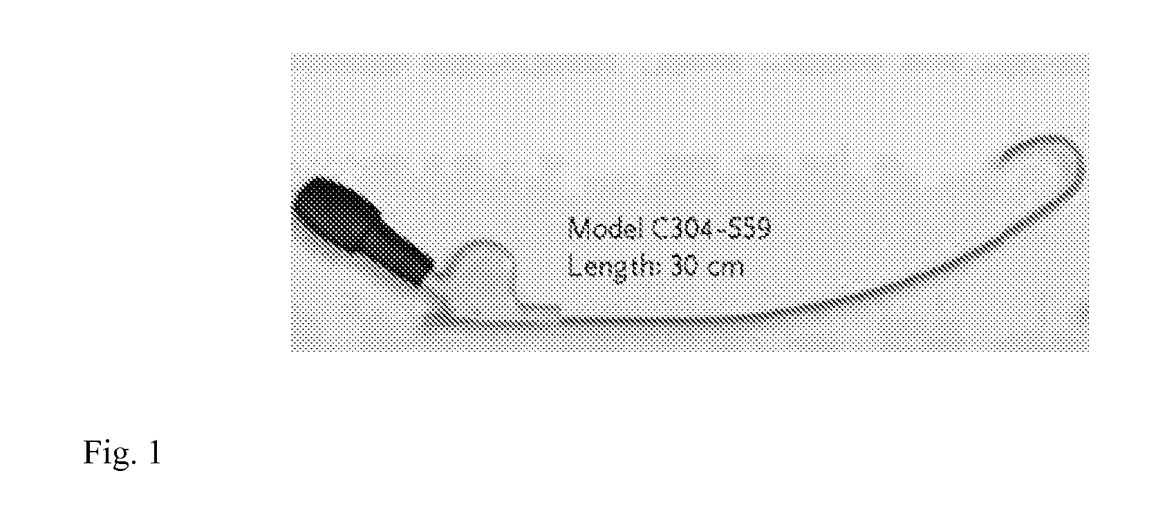
[0083]In an example, a micro-Doppler crystal (such as that used with the Smart Needle Doppler described above) is bonded externally to the tip of (e.g.) a Medtronic SelectSecure lead delivery sheath for pacing purposes, or of a compatible steerable delivery sheath such as those used for ablation. The sheath is then advanced, e.g. using a subxyphoid approach, into the pericardial space. In the case of pacing, when the tip of the sheath reaches an area of the external surface of the LV that is deemed as appearing appropriate for left ventricular pacing, the Doppler probe is used to determine that no blood vessel is in close proximity which blood vessel, in the estimation of the user (for example a physician) is of a size that would be deleterious to the subject's health if adversely affected by the procedure. The lead can then be extended and tested with, or without, fixation to the myocardium. If desired, echocardiographic or other measurements can be made to assess effects on ventri...


PUM


Abstract

A method is provided for placing an epicardial pacing lead onto the epicardium of a heart in a subject. A method is also provided for placing an ablation catheter onto the epicardium of a heart in a subject. Articles of manufacture or machines for use in the methods are also provided.

Description

CROSS-REFERENCE TO RELATED APPLICATIONS[0001]This application claims benefit of U.S. Provisional Application No. 61 / 674,973,filed Jul. 24, 2012, the contents of which are hereby incorporated by reference.BACKGROUND OF THE INVENTION[0002]Throughout this application various publications, patents, patent application publications and books are referred to. Full citations for the publications may be found at the end of the specification. The disclosures of the publications, patents, patent application publications and books are hereby incorporated by reference in their entirety into the subject application to more fully describe the art to which the subject invention pertains.[0003]In patients with heart failure, the electrical impulses that cause squeezing or contraction, and therefore pumping by the lateral part of the left ventricle, are often slower than normal. This results in an asymmetrical or desynchronized left ventricular contraction that compromises the pumping ability of an a...

Claims


Application Information

Patent Timeline
IPC IPC(8): A61B19/00A61B8/08A61B8/02A61B18/02A61N1/362A61N1/05A61B8/06A61B18/14
CPCA61B19/5244A61B8/06A61B8/488A61B18/1492A61B18/02A61N1/362A61B2017/003A61B8/02A61B2018/0212A61B2018/00363A61B2018/00577A61B2019/528A61N1/0587A61B2017/00106A61B2017/0011A61B2018/00863A61B2018/1475A61B2562/0257A61N1/059A61N1/368A61B34/20A61B2090/3784A61N1/3629
Inventor FISHER, JOHN DEVENS
Owner MONTEFIORE MEDICAL CENT INC
Why Eureka
Try Eureka