System and method for performing an ultrasound scan of cellular tissue

a technology of ultrasound and cellular tissue, applied in ultrasonic/sonic/infrasonic image/data processing, tomography, applications, etc., can solve the problems of system time needed to perform the scan, process can still be somewhat slow, and difficulty in identifying abnormalities

Active Publication Date: 2015-09-24
SONOCINE INC
View PDF4 Cites 24 Cited by
  • Summary
  • Abstract
  • Description
  • Claims
  • Application Information

AI Technical Summary

Benefits of technology

[0006]In a first separate aspect of the present invention, a system for performing an ultrasound scan of cellular tissue includes: an ultrasound scanning device including an ultrasound probe, the ultrasound scanning device configured to generate cross-sectional images of the cellular tissue; a probe enclosure in which the ultrasound probe is disposed, wherein a side part of the probe enclosure is formed by a flexible membrane, the probe enclosure is configured to hold an ultrasonic coupling material on the flexible membrane, the flexible membrane and the ultrasonic coupling material being ultrasonically conductive, and a the probe enclosure forms a cavity adjacent the flexible membrane, the cavity being configured to be pressurized above atmospheric pressure; an armature is configured to support the probe enclosure and to move the probe enclosure into a position where the flexible membrane is placed adjacent to and displaced by the cellular tissue; a probe positioning assembly configured to support the ultrasound probe within the probe enclosure and to move the ultrasound probe over the flexible membrane with a head of the ultrasound probe submerged in the ultrasonic coupling material, the ultrasound scanning device generating the cross-sectional images of the cellular tissue as the probe positioning assembly progressively moves the ultrasound probe over the flexible membrane, wherein the probe positioning assembly includes at least one sensor configured to measure a position of the probe within the probe enclosure; and a controller operatively coupled to the probe positioning assembly, wherein the controller is programmed to control movement of the ultrasound probe in response to measurements by the at least one sensor.
[0007]In a second separate aspect of the present invention, a system for performing an ultrasound scan of cellular tissue includes: an ultrasound scanning device including an ultrasound probe, the ultrasound scanning device configured to generate cross-sectional images of the cellular tissue; a probe enclosure in which the ultrasound probe is disposed, wherein a side part of the probe enclosure is formed by a flexible membrane, the probe enclosure is configured to hold an ultrasonic coupling material on the flexible membrane, the flexible membrane and the ultrasonic coupling material being ultrasonically conductive, and a the probe enclosure forms a cavity adjacent the flexible membrane, the cavity being configured to be pressurized above atmospheric pressure; an armature is configured to support the probe enclosure and to move the probe enclosure into a position where the flexible membrane is placed adjacent to and displaced by the cellular tissue; a probe positioning assembly configured to support the ultrasound probe within the probe enclosure and to move the ultrasound probe over the flexible membrane with a head of the ultrasound probe submerged in the ultrasonic coupling material, the ultrasound scanning device generating the cross-sectional images of the cellular tissue as the probe positioning assembly progressively moves the ultrasound probe over the flexible membrane, wherein the probe positioning assembly includes at least one sensor configured to measure a position of the probe within the probe enclosure; and a controller operatively coupled to the probe positioning assembly, wherein the controller is programmed to control movement of the ultrasound probe in response to measurements by the at least one sensor.

Problems solved by technology

One of the limitations of such a system is the amount of time needed to perform the scans.
Even though such a system may automate the acquisition of ultrasound images, the process can still be somewhat slow, especially when compared to other breast tissue screening methods, such as mammography.
Another limitation with present ultrasound systems is that they all rely on the ultrasound probe coming into contact with the surface of the tissue being screened.
This contact causes deformation of the tissue, and deformation of the tissue can cause difficulties for identifying abnormalities when the ultrasound images are being viewed.

Method used


Image

  • System and method for performing an ultrasound scan of cellular tissue
  • System and method for performing an ultrasound scan of cellular tissue
  • System and method for performing an ultrasound scan of cellular tissue

Examples


Embodiment Construction

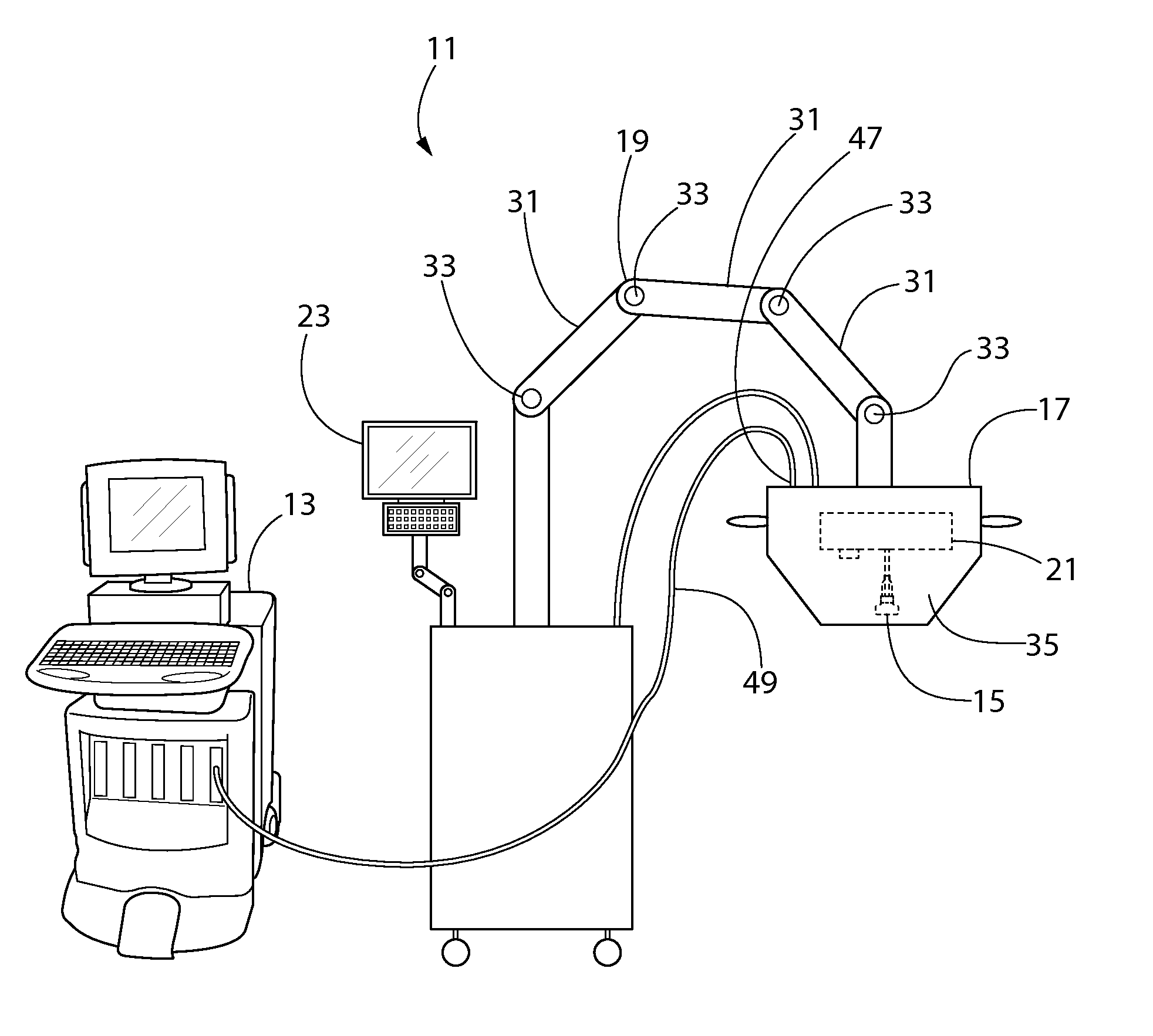
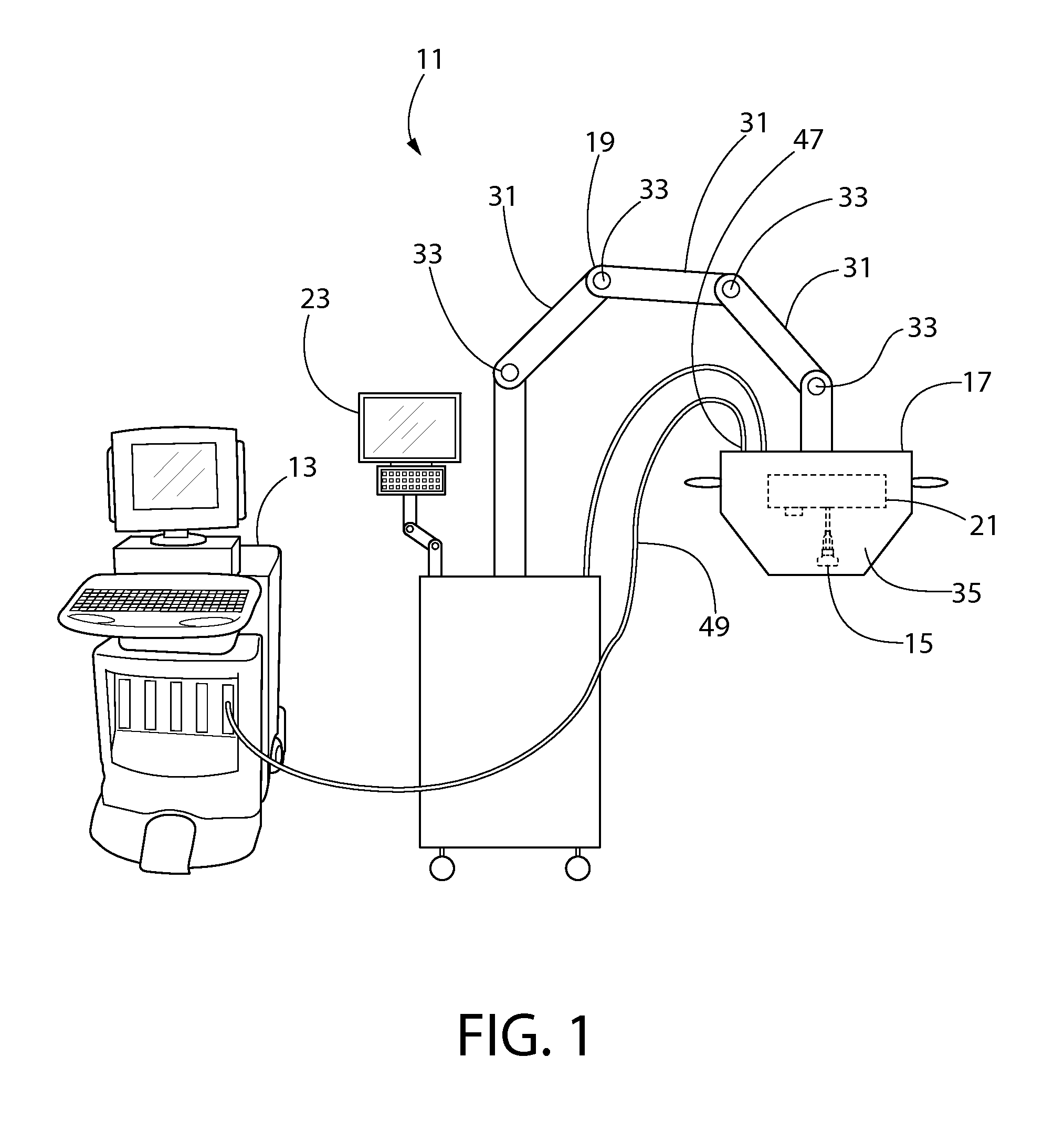
[0026]Turning in detail to the drawings, FIG. 1 illustrates a system 11 for performing an ultrasound scan of cellular tissue. The system 11 includes an ultrasound scanning device 13, which includes an ultrasound probe 15 and is configured to generate a plurality of cross-sectional images of cellular tissue. Unless otherwise indicated in the claims, the ultrasound scanning device 13 may be of a type that is known in the art, which includes a programmable processor, a volatile memory, a non-volatile memory, and programming which is used by the programmable processor to control the ultrasound probe 15, collect ultrasound scan data from the ultrasound probe 15, and to display the ultrasound scan data as images in a desirable format. The processing of ultrasound scan data is described in further detail below.

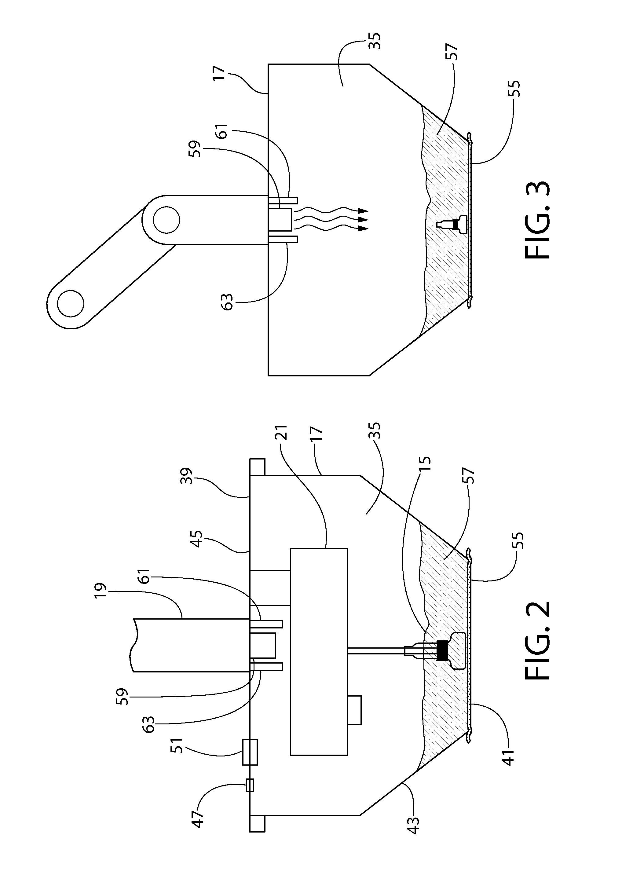
[0027]The ultrasound probe 15 is disposed within the probe enclosure 17, which is supported by an armature 19. The probe enclosure 17 is moved by the armature 19 into a position adja...


PUM


Abstract

A system for performing an ultrasound scan of cellular tissue includes an ultrasound scanning device having an ultrasound probe, to generate cross-sectional images of the cellular tissue. A probe enclosure houses the ultrasound probe, has a bottom formed by a flexible membrane, is configured to hold an ultrasonic coupling material, and forms a pressurizable cavity adjacent the flexible membrane. An armature supports the probe enclosure and is configured to move the probe enclosure into a position where the flexible membrane is placed adjacent to and displaced by the cellular tissue. A probe positioning assembly supports the ultrasound probe within the probe enclosure and moves the ultrasound probe over the flexible membrane with a head of the ultrasound probe submerged in the ultrasonic coupling material, the ultrasound scanning device generating the cross-sectional images of the cellular tissue as the probe positioning assembly progressively moves the ultrasound probe over the flexible membrane.

Description

CROSS REFERENCE TO RELATED APPLICATIONS[0001]Priority is claimed to U.S. provisional patent application No. 61 / 968,712, filed Mar. 21, 2014, the disclosure of which is incorporated herein by reference in its entirety.FIELD OF THE INVENTION[0002]The field of the present invention relates to ultrasonic scanning and diagnostics for cellular tissue.BACKGROUND OF THE INVENTION[0003]Ultrasound as an independent screening tool for diagnosing cancers, particularly in breast tissue, is becoming more widely recognized and accepted within the medical community. Historically, in the context of breast exams and breast cancers, ultrasound was first used to diagnose breast cancers if the location of the abnormality is first discovered by another modality, such as mammography or physical examination. Recently, ultrasound as a stand-alone diagnostic tool for breast cancers has become more widely accepted based on the development of technology that enables ultrasound to be used to screen the entire b...

Claims


Application Information

Patent Timeline
Patent Type & Authority Applications(United States)
IPC IPC(8): A61B8/00A61B8/08A61B8/14
CPCA61B8/4444A61B8/14A61B8/40A61B8/42A61B8/0825A61B8/5207A61B8/4218A61B8/4209A61B8/4245A61B6/0407
Inventor KELLY, KEVIN M.
Owner SONOCINE INC
Why Eureka
Try Eureka